. 2015 Sep 22;11(9):e1004508.
doi: 10.1371/journal.pcbi.1004508. eCollection 2015.

Systematic Prediction of Scaffold Proteins Reveals New Design Principles in Scaffold-Mediated Signal Transduction

Affiliations

Systematic Prediction of Scaffold Proteins Reveals New Design Principles in Scaffold-Mediated Signal Transduction

Jianfei Hu et al. PLoS Comput Biol. .

Abstract

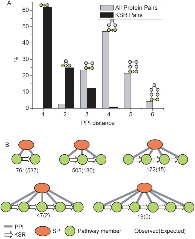
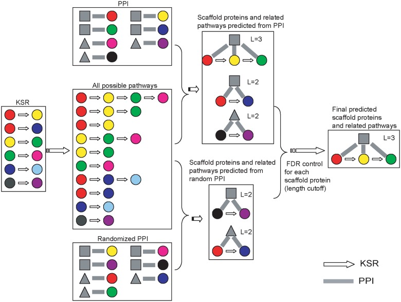
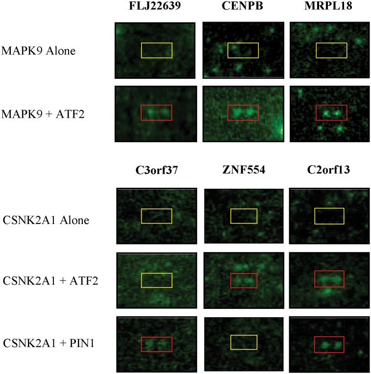
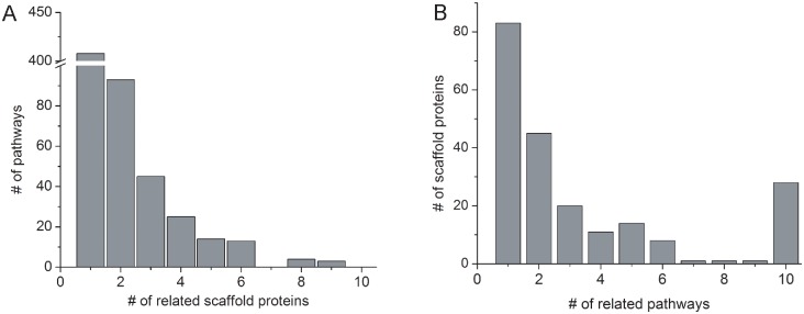
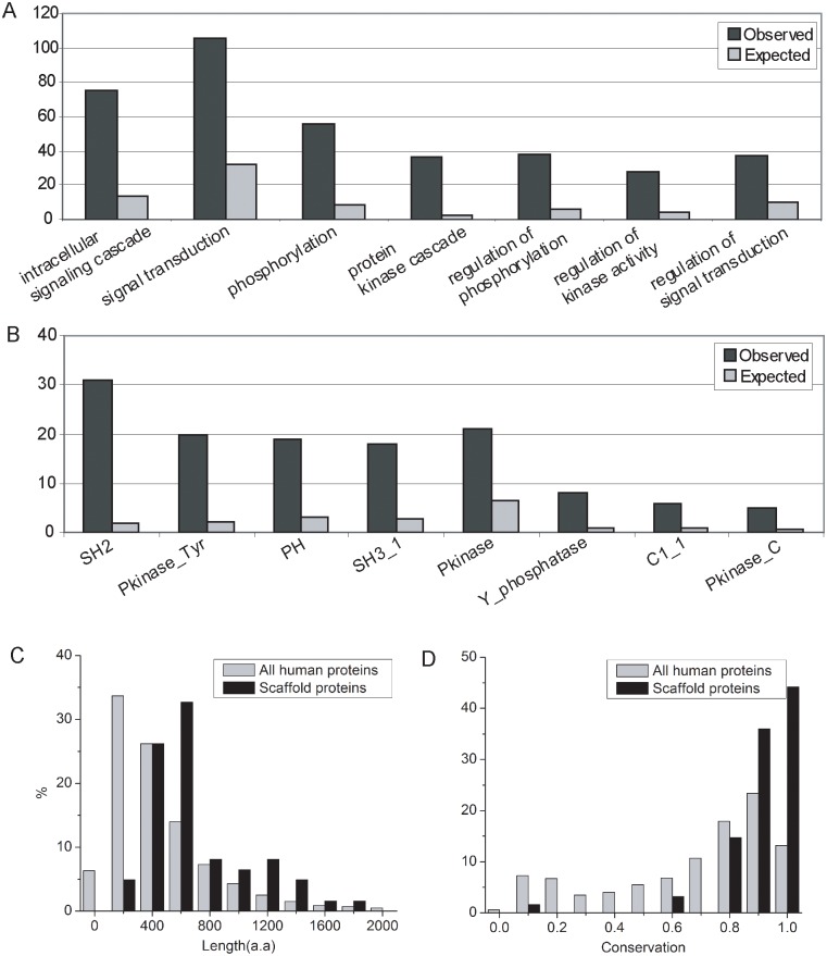
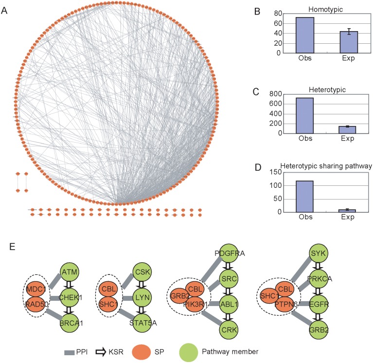
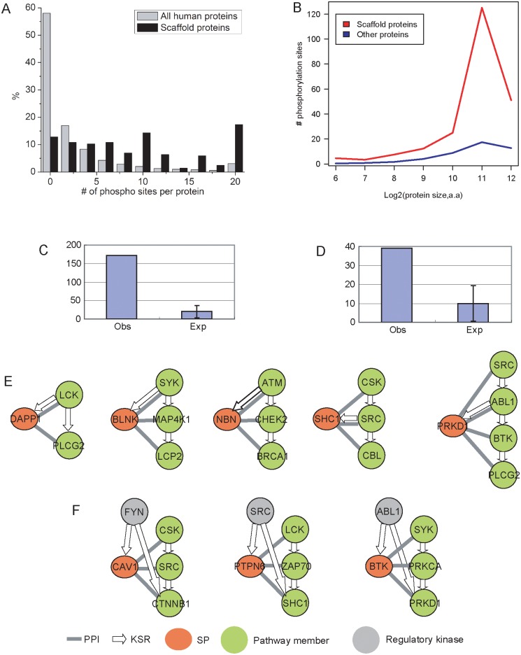
Scaffold proteins play a crucial role in facilitating signal transduction in eukaryotes by bringing together multiple signaling components. In this study, we performed a systematic analysis of scaffold proteins in signal transduction by integrating protein-protein interaction and kinase-substrate relationship networks. We predicted 212 scaffold proteins that are involved in 605 distinct signaling pathways. The computational prediction was validated using a protein microarray-based approach. The predicted scaffold proteins showed several interesting characteristics, as we expected from the functionality of scaffold proteins. We found that the scaffold proteins are likely to interact with each other, which is consistent with previous finding that scaffold proteins tend to form homodimers and heterodimers. Interestingly, a single scaffold protein can be involved in multiple signaling pathways by interacting with other scaffold protein partners. Furthermore, we propose two possible regulatory mechanisms by which the activity of scaffold proteins is coordinated with their associated pathways through phosphorylation process.


Conflict of interest statement

The authors have declared that no competing interests exist.

Figures

Fig 1
Fig 1. Scaffold proteins are widespread in signaling networks.
(A) PPI distance of KSR pairs and all human protein pairs. The PPI distance of a protein pair is defined as the shortest distance of the two proteins in PPI network. KSR pairs are significantly enriched in PPI distance = 2. In fact, 24.9% of KSR pairs have PPI distance of 2, while only 2.7% of all human protein pairs have the same PPI distance. (B) Network motifs in which one protein interacts with a series of proteins and these proteins form a cascade via KSRs. These network motifs are enriched, suggesting that scaffold proteins are widespread in signaling pathways.
Fig 2
Fig 2. Strategy to predict scaffold proteins.
For each potential scaffold protein, we corrected the effect of interaction degree of the protein and the length of associated pathways. We utilized the randomized PPI to assess the significance of a predicted scaffold protein. The random PPI keep the same PPI degree for each protein by randomly selecting two PPI pairs and changing their partners.
Fig 3
Fig 3. Experimental validations for CSNK2A1 and MAPK9.
A human proteome microarray, comprised of 17,000 individually purified human proteins in full-length, was used to perform phosphorylation reactions with CKII (CSNK2A1) and JNK2 (MAPK9) in the presence or absence of their predicted scaffold proteins, ATF2 and PIN1. Phosphorylation signals were detected by exposure of the human proteome microarrays to X-ray film. Positive hits in red boxes were identified by visual inspection.
Fig 4
Fig 4. Specificity of scaffold proteins and pathways.
(A) Number of pathways related to scaffold proteins. 408 pathways (67.4%) are found to be associated with only one scaffold protein. (B) Number of scaffold proteins related to pathways. Specifically, 83 scaffold proteins are associated with only one pathways, while 28 scaffold proteins are related to >10 pathways.
Fig 5
Fig 5. Characterization of scaffold proteins.
(A) Enriched GO terms for scaffold proteins. (B) Enriched protein domains defined by Pfam in scaffold proteins. The GO and Pfam terms are sorted increasingly from left to right by p-value. (C) Distribution of protein lengths. (D) Distribution of evolutionary conservation.
Fig 6
Fig 6. Interactions between scaffold proteins.
(A) Overall PPI interaction networks between scaffold proteins. (B) Number of homotypic interactions among scaffold proteins. (C) Number of heterotypic interactions among scaffold proteins. (D) Number of heterotypic interactions that are associated with the same pathways. (E) Examples of scaffold protein complexes that are associated with signaling pathways. The scaffold proteins in a complex interact each other, and all of them interact with each member in the associated pathway.
Fig 7
Fig 7. Possible regulatory mechanisms of scaffold proteins.
(A) Distribution of MS/MS determined phosphorylation site numbers per protein. Predicted scaffold proteins have 12 sites on average. As a contrast, proteins in human proteome only have 2 sites on average. (B) The high number of phosphorylation sites in scaffold proteins is not due to larger protein sizes. If we compared the number of sites between scaffold proteins and the proteins with similar sizes, the similar observation was made. One possible regulatory mechanism for scaffold proteins is that one kinase member in the pathway phosphorylates the scaffold protein. The number of such cases is shown in (C). The examples are shown in (E). The second possible mechanism is that one upstream kinase phosphorylates both scaffold protein and one member in the pathways. The number of such cases is shown in (D). The examples are shown in (F).

References

    1. Linding R, Jensen LJ, Ostheimer GJ, van Vugt MA, Jorgensen C, et al. (2007) Systematic discovery of in vivo phosphorylation networks. Cell 129: 1415–1426. - PMC - PubMed
    1. Fiedler D, Braberg H, Mehta M, Chechik G, Cagney G, et al. (2009) Functional organization of the S. cerevisiae phosphorylation network. Cell 136: 952–963. 10.1016/j.cell.2008.12.039 - DOI - PMC - PubMed
    1. Newman RH, Hu J, Rho HS, Xie Z, Woodard C, et al. (2013) Construction of human activity-based phosphorylation networks. Mol Syst Biol 9: 655 10.1038/msb.2013.12 - DOI - PMC - PubMed
    1. Hu J, Rho HS, Newman RH, Hwang W, Neiswinger J, et al. (2014) Global analysis of phosphorylation networks in humans. Biochim Biophys Acta 1844: 224–231. 10.1016/j.bbapap.2013.03.009 - DOI - PMC - PubMed
    1. Hu J, Rho HS, Newman RH, Zhang J, Zhu H, et al. (2014) PhosphoNetworks: a database for human phosphorylation networks. Bioinformatics 30: 141–142. 10.1093/bioinformatics/btt627 - DOI - PMC - PubMed

Publication types

LinkOut - more resources