. 2015 Nov 1;142(21):3686-91.
doi: 10.1242/dev.128942. Epub 2015 Sep 22.

Phenotypic analysis of mice completely lacking netrin 1

Affiliations

Phenotypic analysis of mice completely lacking netrin 1

Andrea R Yung et al. Development. .

Abstract

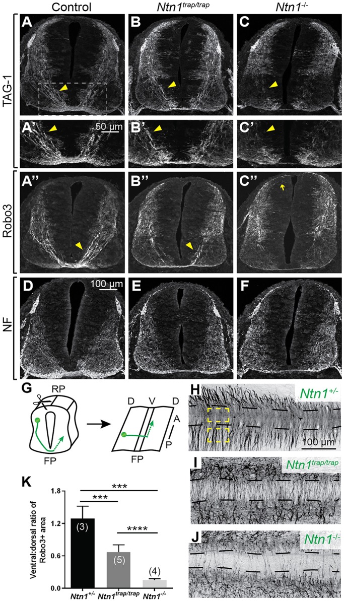
Netrin 1 (Ntn1) is a multifunctional guidance cue expressed in the ventricular zone and floor plate of the embryonic neural tube. Although Ntn1 is best known for acting as an axon guidance cue through Dcc and neogenin receptors, it is also thought to regulate neuronal survival and blood vessel development through Unc5 family receptors. However, the Ntn1 gene trap mutant mouse does not display all the phenotypes predicted from in vitro assays or analyses of mice lacking predicted receptors. Since the gene trap strain still produces wild-type Ntn1 protein, it is unclear whether the absence of phenotypes reflects the activity of alternative cues or of residual Ntn1. To resolve the full contribution of Ntn1 to development, we generated a null allele of Ntn1 and re-examined tissues exhibiting phenotypic discrepancies between receptor mutants and Ntn1 hypomorphs. We found that in Ntn1 null animals commissural axons rarely cross the midline, resulting in a strongly enhanced phenotype relative to Ntn1 hypomorphs, which retain many axons with normal trajectories. Thus, low levels of Ntn1 can account for persistent attraction to the midline in hypomorphs. By contrast, Ntn1 null mice do not show all of the phenotypes reported for Unc5 receptor mutants, indicating that Ntn1 is not necessarily the dominant ligand for Unc5 family members in vivo and ruling out primary roles in survival or angiogenesis.

Keywords: Axon guidance; Commissural neurons; Netrin-1.


Conflict of interest statement

Competing interests

The authors declare no competing or financial interests.

Figures

Fig. 1.
Fig. 1.
Generation of the Ntn1 null mouse. (A) Map of the wild-type and floxed Ntn1 loci with GenBank annotations. loxP sites flank exon 2; its protein product (yellow, domain VI; blue, domain V) is delineated by dashed lines. (B) Western blots of E11.5 head lysate show residual protein in Ntn1trap/trap mutants but no detectable protein in the newly generated Ntn1−/− mutants. Loading controls (actin) were obtained from a shorter exposure of the same gel.
Fig. 2.
Fig. 2.
The Ntn1trap/trap commissural phenotype is enhanced in Ntn1−/− mutants. (A-C″) Low (A-C,A″-C″) and high (A′-C′) magnification views of E11.5 spinal cord sections stained for TAG-1 and Robo3 reveal that fewer commissural axons (arrowheads) cross the midline in Ntn1−/− animals (C) compared with controls (A) and Ntn1trap/trap hypomorphs (B), with some axons projecting dorsally (arrow). (D-F) Neurofilament (NF) stains show grossly normal organization of the spinal cord in E11.5 null mutants. (G-J) Robo3 staining of open-book preparations of E11.5 spinal cords (G) show that fewer axons cross the midline (dashed lines) in Ntn1trap/trap animals (I) compared with controls (H). This phenotype is more severe in Ntn1−/− animals (J). (K) Quantification of the midline crossing phenotypes illustrated in H-J. Yellow boxes in H indicate the dorsal and ventral areas quantified. RP, roof plate; FP, floor plate; D, dorsal; V, ventral. ****P<0.0001, ***P<0.001; Mann–Whitney test. Error bars indicate s.d.
Fig. 3.
Fig. 3.
Ntn1−/− mutants maintain expression of other guidance cues and their receptors. (A-H) In situ hybridization of E11.5 spinal cord sections; dashed lines indicate the ventral edge. Shh (A,B), Vegf (C,D), Slit1 (E,F) and Slit2 (G,H) are expressed at the floor plate of mutant animals, as in wild-type (WT) controls. (I) Quantified western blots for Dcc, neogenin, Flk1 and Boc show similar, or upregulated, levels of these receptors in wild-type and null animals. *P<0.05; Student's t-test. Error bars indicate s.d. (J-K′) Immunostaining (J,J′) and in situ hybridization (K,K′) confirm that Dcc and Boc expression is preserved in E11.5 mutant spinal cords.
Fig. 4.
Fig. 4.
Ntn1−/− mutants do not display many known Unc5 receptor mutant phenotypes. (A-D′) Colorimetric and fluorescent wholemount neurofilament stains show comparable sensory and motor projections in wild-type (A) and Ntn1 null (B) E11.5 embryos, including normal trochlear (IV) nerve trajectories (C,D) with an intact dorsal decussation (C′,D′; arrowheads indicate trochlear nerve on either side). Cranial nerves III, IV and V are indicated. (E-F) Islet1/2-positive trochlear nuclei retain their normal position relative to the midline in mutant E12.5 coronal hindbrain sections, as quantified in F. (G-J) SACMN axons normally form a smooth, hook-shaped nerve (white arrow), but both null (H,I) and hypomorphic (J) mutants show variable defects, ranging from a few axons (yellow arrowheads) to whole bundles of axons (yellow arrows) wandering away from the nerve at many positions. (K-L) Islet1 immunostains show no change in the number of motor neurons in the spinal cord of E13.5 wild-type and null animals, as quantified in L. (M-N) Immunostaining for PECAM in E12.5 hindbrain sections show no change in blood vessel coverage, as quantified in N. n.s., not significant (F, P=0.474; L, P=0.445; N, P=0.480; Mann–Whitney test). Error bars indicate s.d.

References

    1. Bin J. M., Rajasekharan S., Kuhlmann T., Hanes I., Marcal N., Han D., Rodrigues S. P., Leong S. Y., Newcombe J., Antel J. P. et al. (2013). Full-length and fragmented netrin-1 in multiple sclerosis plaques are inhibitors of oligodendrocyte precursor cell migration. Am. J. Pathol. 183, 673-680. 10.1016/j.ajpath.2013.06.004 - DOI - PubMed
    1. Bin J. M., Han D., Lai Wing Sun K., Croteau L.-P., Dumontier E., Cloutier J.-F., Kania A. and Kennedy T. E. (2015). Complete loss of Netrin-1 results in embryonic lethality and severe axon guidance defects without increased neural cell death. Cell Rep. 12, 1099-1106. 10.1016/j.celrep.2015.07.028 - DOI - PubMed
    1. Brunet I., Gordon E., Han J., Cristofaro B., Broqueres-You D., Liu C., Bouvrée K., Zhang J., del Toro R., Mathivet T. et al. (2014). Netrin-1 controls sympathetic arterial innervation. J. Clin. Invest. 124, 3230-3240. 10.1172/JCI75181 - DOI - PMC - PubMed
    1. Burgess R. W., Jucius T. J. and Ackerman S. L. (2006). Motor axon guidance of the mammalian trochlear and phrenic nerves: dependence on the netrin receptor Unc5c and modifier loci. J. Neurosci. 26, 5756-5766. 10.1523/JNEUROSCI.0736-06.2006 - DOI - PMC - PubMed
    1. Charron F., Stein E., Jeong J., McMahon A. P. and Tessier-Lavigne M. (2003). The morphogen sonic hedgehog is an axonal chemoattractant that collaborates with netrin-1 in midline axon guidance. Cell 113, 11-23. 10.1016/S0092-8674(03)00199-5 - DOI - PubMed

Publication types

LinkOut - more resources