. 2015 Sep 29;6(29):26615-32.
doi: 10.18632/oncotarget.5666.

Radiation-induced lung damage promotes breast cancer lung-metastasis through CXCR4 signaling

Affiliations

Radiation-induced lung damage promotes breast cancer lung-metastasis through CXCR4 signaling

Lynn Feys et al. Oncotarget. .

Abstract

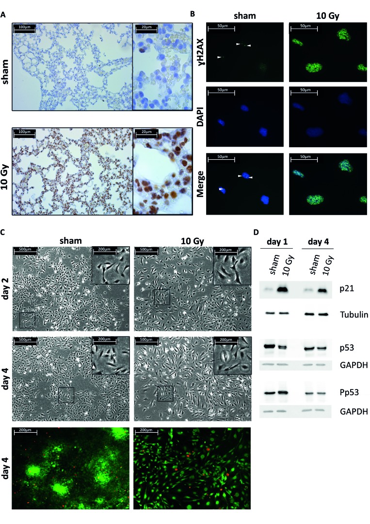
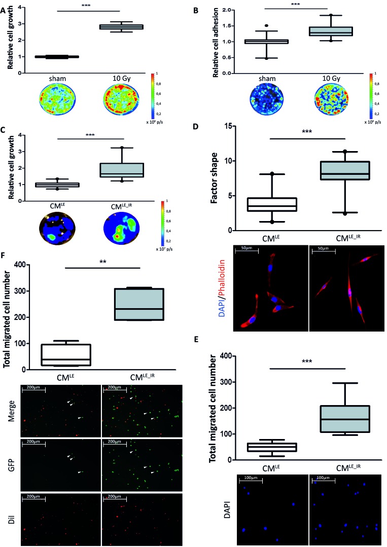
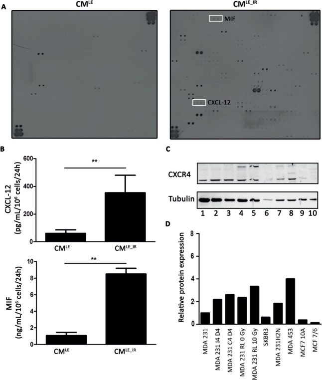
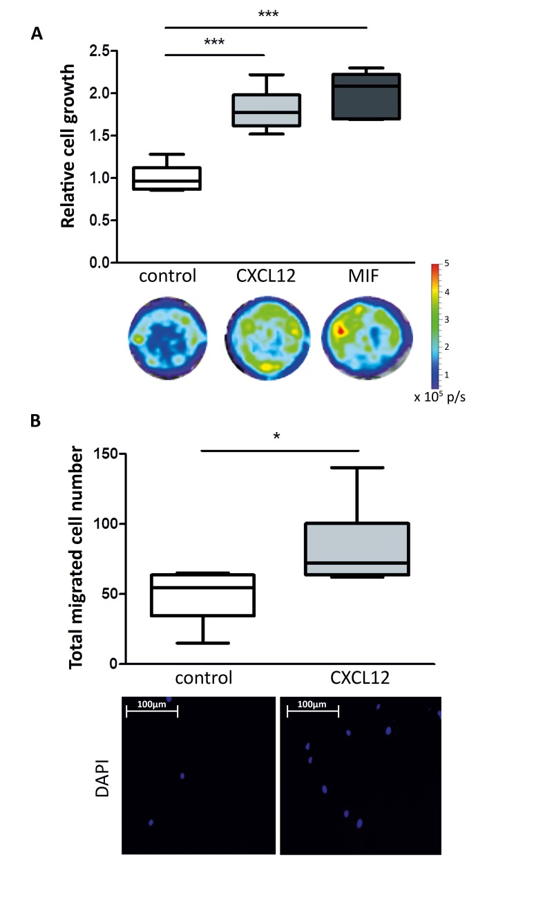
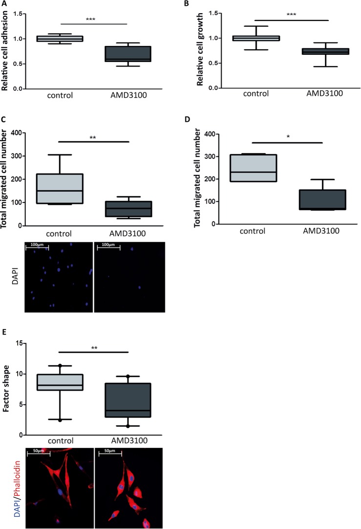
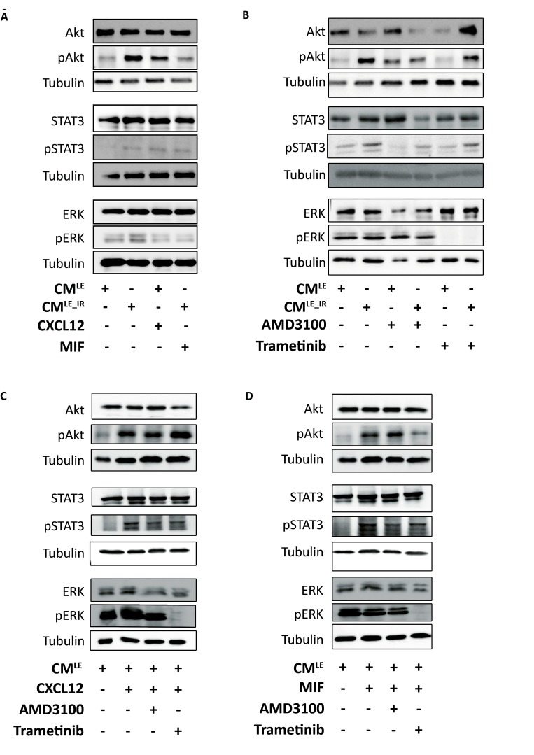
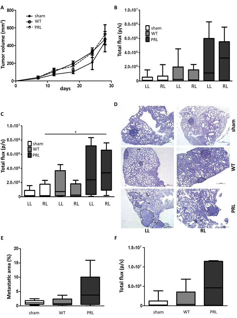
Radiotherapy is a mainstay in the postoperative treatment of breast cancer as it reduces the risks of local recurrence and mortality after both conservative surgery and mastectomy. Despite recent efforts to decrease irradiation volumes through accelerated partial irradiation techniques, late cardiac and pulmonary toxicity still occurs after breast irradiation. The importance of this pulmonary injury towards lung metastasis is unclear. Preirradiation of lung epithelial cells induces DNA damage, p53 activation and a secretome enriched in the chemokines SDF-1/CXCL12 and MIF. Irradiated lung epithelial cells stimulate adhesion, spreading, growth, and (transendothelial) migration of human MDA-MB-231 and murine 4T1 breast cancer cells. These metastasis-associated cellular activities were largely mimicked by recombinant CXCL12 and MIF. Moreover, an allosteric inhibitor of the CXCR4 receptor prevented the metastasis-associated cellular activities stimulated by the secretome of irradiated lung epithelial cells. Furthermore, partial (10%) irradiation of the right lung significantly stimulated breast cancer lung-specific metastasis in the syngeneic, orthotopic 4T1 breast cancer model.Our results warrant further investigation of the potential pro-metastatic effects of radiation and indicate the need to develop efficient drugs that will be successful in combination with radiotherapy to prevent therapy-induced spread of cancer cells.

Keywords: AMD3100; CXCL12; MIF; radiotherapy; triple-negative breast cancer.


Conflict of interest statement

CONFLICTS OF INTERESTS

There are no conflicts of interests.

Figures

Figure 1
Figure 1. Lung epithelial cells radiation response and senescence markers
A. Immunohistochemical (IHC) staining of γH2AX foci using an immunoenzymatic DAB staining method (brown color) combined with a haematoxylin counterstaining in sham or 10 Gy irradiated mouse lung tissue. B. Immunocytochemical (ICC) staining of γH2AX foci (Alexa488 labeled secondary antibody, green color) combined with a DAPI nuclear counterstaining (blue color) in sham or 10 Gy irradiated Beas-2B lung epithelial cells. C. Upper 4 panels, phase contrast micrographs of Beas-2B lung epithelial cells two or four days post sham or 10 Gy irradiation. The 10 Gy condition shows less dense cell culture, a more spread cell morphology with enlarged nuclei and increased cytoplasmic surface area. Lower 2 panels, live/dead - viability/cytotoxicity test. Assay shows live cells as green and dead cells as red. Four days after single irradiation dose of 10 Gy shows no increase of Beas-2B cell death. D. Western blot (WB) analysis of p53 and p21 on total cell lysates from Beas-2B cells treated with single-fraction 10 Gy or sham. Total p53 expression is unchanged after irradiation but increase in p53 phosphorylation is observed at day 1 after treatment and normalizes at day 4. Total expression of p21 is increased until day 4. GAPDH and tubulin are used as loading control.
Figure 2
Figure 2. Impact of irradiated lung epithelial cells on breast cancer cell adhesion and growth
A. Box plots illustrating the relative cell growth of MDA-MB-231GFP_luc cells. Co-culture of breast cancer with irradiated Beas-2B cells increases relative cell growth compared to co-culture with sham treated Beas-2B cells. Quantification by bioluminescent imaging after 4 days incubation. Data is represented as relative fold change compared with the corresponding control value. MDA-MB-231GFP_luc: n = 6; ***, P < 0.001 (Unpaired t-test with Welch's correction). B. Box plots illustrating the relative cell adhesion of MDA-MB-231GFP_luc cells. Relative Adhesion of breast cancer cells to irradiated Beas-2B cell monolayer is increased compared to sham treated Beas-2B cell monolayer. Quantification by bioluminescent imaging after 24 hours incubation. Data is represented as relative fold change compared with the corresponding control value. MDA-MB-231GFP_luc: n = 15; ***, P < 0.0001; circle = outlier (Unpaired t-test with Welch's correction). Impact of CMLE_IR on breast cancer cell morphology, growth, migration and extravasation. C. Box plots illustrating the relative cell growth of MDA-MB-231GFP_luc cells. Treatment of breast cancer cells with CMLE_IR increases relative cell growth compared to cells treated with CMLE. Quantification by bioluminescent imaging after 4 days incubation. Data is represented as relative fold change compared with the corresponding control value (CMLE). MDA-MB-231GFP_luc: n = 21; ***, P < 0.001 (Mann-Whitney U). D. Box plots illustrating the extent of cell spreading of MDA-MB-231GFP_luc cells in CMLE_IR versus CMLE conditions, as quantified by factor shape (upper panel). Treatment with CMLE_IR showed enhanced cell spreading, corresponding with the significantly altered cell shapes (lower panel). Fluorescence microscopy images of cells double stained with phalloidin for actin filaments (red) and DAPI counterstaining for nuclei (blue) after 4 days of incubation with CMLE_IR versus CMLE. n = 20; ***, P < 0.001 (Mann-Whitney U). E. Box plots illustrating total migrated cell number of MDA-MB-231GFP_luc cells in CMLE_IR versus CMLE conditions (upper panel). Nuclei of migrated cells were stained blue by DAPI (lower panel). n = 12; ***, P < 0.001 (Unpaired t-test with Welch's correction). F. Box plots illustrating total migrated cell number of MDA-MB-231GFP_luc cells through endothelial monolayer in CMLE_IR versus CMLE conditions (upper panel). CMLE_IR enhances breast cancer cell extravasation significantly. Endothelial cells were stained red with Vibrant DiI. Extravasated MDA-MB-231GFP_luc cells are green (lower panel). n = 6; **, P = 0.0022 (Mann-Whitney U). Arrowheads indicating GFP positive migrated breast cancer cells.
Figure 3
Figure 3. Increased secretion of CXCL12 and MIF by irradiated lung epithelial cells
A. Cytokine array identifies enhanced presence of CXCL12 and MIF in CMLE_IR compared to CMLE. B. ELISA analysis of CXCL12 (upper panel) and MIF (lower panel) on CMLE_IR versus CMLE. For each condition 2 different samples were used in triplicate. Values are presented as the mean ± SD. CXCL12: **, P = 0.010. MIF: **, P = 0.004 (Mann-Whitney U). C. Western blot analysis of CXCR4 expression on total lysate of breast cancer cells. Lane 1-5, represents MDA-MB-231GFP_luc (invasive cell line) in different conditions (MDA 231: parental MDA-MB-231GFP_luc; MDA 231 I4 D4: MDA 231 cells exposed to CMLE_IR for 4 days; MDA 231 C4 D4: MDA 231 cells exposed to CMLE for 4 days; MDA 231 RL 0 Gy: MDA 231 isolated from non-irradiated mouse lung; MDA 231 RL 10 Gy: MDA 231 isolated from irradiated mouse lung. Lane 6: SKBR3 (non-invasive cell line). Lane 7: MDA-MB-231H2N (invasive cell line). Lane 8: MDA-MB-453 (invasive cell line). Lane 9: MCF 10A (non-invasive cell line). Lane 10: MCF7/6 (non-invasive cell line). D. Quantitative analysis CXCR4 protein expression level. Western blot analysis of CXCR4 protein expression in different breast cancer cell lines after tubulin normalization. Protein levels relative to control, MDA-MB-231GFP_luc.
Figure 4
Figure 4. Effect of recombinant CXCL12 and MIF on breast cancer cell growth and migration
A. Box plots illustrating the relative cell growth of MDA-MB-231GFP_luc cells. Treatment of breast cancer cells with CXCL12 (50 ng/mL) or MIF (50 ng/mL) increases relative cell growth compared to control. Quantification by bioluminescent imaging after 4 days incubation. Data is represented as relative fold change compared with the corresponding control value (CMLE). n = 6; ***, P < 0.001 (Unpaired t-test). B. Box plots illustrating difference in total migrated cell number of MDA-MB-231GFP_luc cells treated with CXCL12 (50 ng/mL) (upper panel). Nuclei of migrated cells were stained blue with DAPI (lower panel). n = 6; *, P = 0.038 (Unpaired t-test).
Figure 5
Figure 5. Effect of an allosteric CXCR4 inhibitor on breast cancer cell growth and migration
A. Box plots illustrating the relative cell adhesion of MDA-MB-231GFP_luc cells. Treatment of breast cancer cells with AMD3100 (10 μM) decreases relative cell adhesion to irradiated Beas-2B cell monolayer. n = 12; ***, P < 0.001(Unpaired t-test with Welch's correction). B. Box plots illustrating the relative cell growth of MDA-MB-231GFP_luc cells. Treatment of breast cancer cells with AMD3100 (10 μM) decreases relative cell growth in presence of CMLE-IR. (Unpaired t-test with Welch's correction). n = 15; ***, P < 0.001 (Unpaired t-test). In C. and D., quantification by bioluminescent imaging after 4 days incubation. Data is represented as relative fold change compared with the corresponding control value. C. Box plots illustrating impact of AMD3100 (10 μM) on total migrated cell number of MDA-MB-231GFP_luc cells stimulated by CMLE-IR. Nuclei were stained blue with DAPI (lower panel). n = 9; **, P = 0.009 (Unpaired t-test with Welch's correction). D. Box plots illustrating total transendothelial migrated cell number of MDA-MB-231GFP_luc cells stimulated by CMLE-IR in presence of AMD3100 (10 μM) or control. n = 6; *, P = 0.017 (Mann-Whitney U). E. Box plots illustrating the extent of CMLE-IR -induced cell spreading of MDA-MB-231GFP_luc cells treated with AMD3100 (10 μM) versus control, as quantified by factor shape (upper panel). Fluorescence microscopy images of cells double stained with phalloidin for actin filaments (red) and DAPI counterstaining for nuclei (blue) (lower panel). n = 20; **, P = 0.004 (Mann-Whitney U).
Figure 6
Figure 6. Paracrine activation of ERK, Akt and STAT3 in breast cancer cells by CMLE_IR and recombinant CXCL12
A.-D.. Western blot analysis of pERK, pAkt and pSTAT3 in MDA-MB-231GFP_luc cells treated with recombinant CXCL12 (50 ng/mL) or MIF (50 ng/mL) A. CMLE, CMLE_IR, in presence of AMD3100 (10 μM) or Trametinib (50 nM) B. recombinant CXCL12 (50 ng/mL) C. or MIF (50 ng/mL) D. in presence of AMD3100 (10 μM) or Trametinib (50 nM) Tubulin is used as loading control.
Figure 7
Figure 7. Impact of partial lung irradiation on lung-specific breast cancer metastasis in a syngeneic mouse model
A. 4 week old BALB/c female mice were injected orthotopically with 1 × 10⁶ 4T1_luc cells in 0.1 mL of serum free DMEM with 50% Matrigel. Tumor formation was monitored for 28 days by caliper measurement. Tumor volumes were measured as indicated. Sham and WT IR: n = 6 and PRL: n = 5. B.-C. & F. 28 days after implantation of the cells, mice were sacrificed and total lung metastasis was quantified by bioluminescent imaging. Lung were quantified separately (B & C) or as one (F). Sham and WT: LL and RL, n = 6 and PRL: LL and RL, n = 5 (B). Only lungs containing metastasis were quantified, showing significant increase in signal in right lung of PRL mice. Sham: LL, n = 5; RL, n = 4; WT: LL, n = 4; RL, n = 5; PRL: LL, n = 4; RL, n = 4; *; P = 0.043 (Man-Whitney U test) (C). D. H&E staining of lungs from indicated groups. The metastatic areas are encircled (black). E. Quantification of the percentage of lung metastatic area calculated per mouse. Sham and WT: n = 6 and PRL: n = 5.

References

    1. Goldhirsch A, Wood WC, Coates AS, Gelber RD, Thürlimann B, Senn HJ. Strategies for subtypes--dealing with the diversity of breast cancer: highlights of the St. Gallen International Expert Consensus on the Primary Therapy of Early Breast Cancer 2011. Ann Oncol. 2011;22:1736–1747. - PMC - PubMed
    1. McGale P, Taylor C, Correa C, Cutter D, Duane F, Ewertz M, Gray R, Mannu G, Peto R, Whelan T, Wang Y, Wang Z, Darby S. Effect of radiotherapy after mastectomy and axillary surgery on 10-year recurrence and 20-year breast cancer mortality: meta-analysis of individual patient data for 8135 women in 22 randomised trials. The Lancet. 2014;383:2127–2135. - PMC - PubMed
    1. Moran MS. Radiation therapy in the locoregional treatment of triple-negative breast cancer. The Lancet Oncology. 2015;16:113–122. - PubMed
    1. Clarke M, Collins R, Darby S, Davies C, Elphinstone P, Evans E, Godwin J, Gray R, Hicks C, James S, MacKinnon E, McGale P, McHugh T, Peto R, Taylor C, Wang Y, et al. Effects of radiotherapy and of differences in the extent of surgery for early breast cancer on local recurrence and 15-year survival: an overview of the randomised trials. The Lancet. 2005;366:2087–2106. - PubMed
    1. Darby S, McGale P, Correa C, Taylor C, Arriagada R, Clarke M, Cutter D, Davies C, Ewertz M, Godwin J, Gray R, Pierce L, Whelan T, et al. Early Breast Cancer Trialists' Collaborative Group (EBCTCG). Effect of radiotherapy after breast-conserving surgery on 10-year recurrence and 15-year breast cancer death: meta-analysis of individual patient data for 10 801 women in 17 randomised trials. The Lancet. 2011;378:1707–1716. - PMC - PubMed

Publication types

MeSH terms

Substances