. 2016 Jan;30(1):336-47.
doi: 10.1096/fj.15-277095. Epub 2015 Sep 22.

LRRK2 autophosphorylation enhances its GTPase activity

Affiliations

LRRK2 autophosphorylation enhances its GTPase activity

Zhiyong Liu et al. FASEB J. 2016 Jan.

Abstract

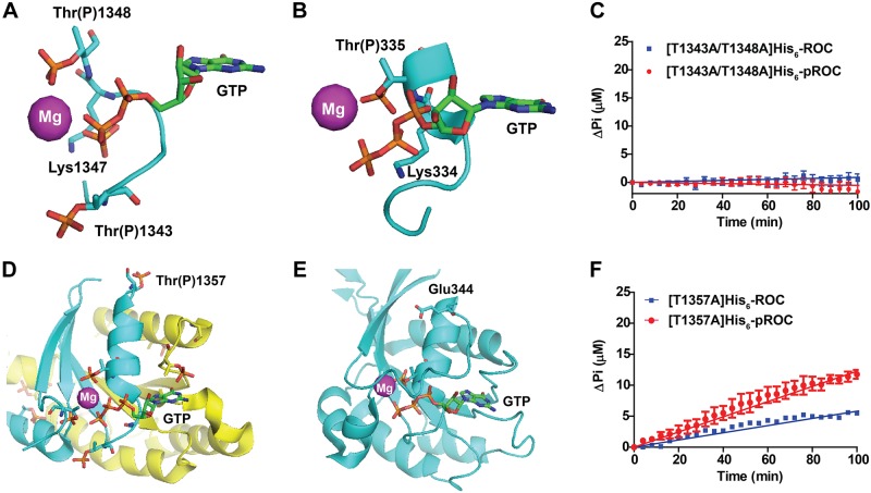
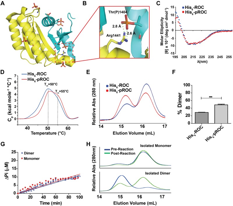
The leucine-rich repeat kinase (LRRK)-2 protein contains nonoverlapping GTPase and kinase domains, and mutation in either domain can cause Parkinson disease. GTPase proteins are critical upstream modulators of many effector protein kinases. In LRRK2, this paradigm may be reversed, as the kinase domain phosphorylates its own GTPase domain. In this study, we found that the ameba LRRK2 ortholog ROCO4 phosphorylates the GTPase domain [termed Ras-of-complex (ROC) domain in this family] of human LRRK2 on the same residues as the human LRRK2 kinase. Phosphorylation of ROC enhances its rate of GTP hydrolysis [from kcat (catalytic constant) 0.007 to 0.016 min(-1)], without affecting GTP or GDP dissociation kinetics [koff = 0.093 and 0.148 min(-1) for GTP and GDP, respectively). Phosphorylation also promotes the formation of ROC dimers, although GTPase activity appears to be equivalent between purified dimers and monomers. Modeling experiments show that phosphorylation induces conformational changes at the critical p-loop structure. Finally, ROC appears to be one of many GTPases phosphorylated in p-loop residues, as revealed by alignment of LRRK2 autophosphorylation sites with GTPases annotated in the phosphoproteome database. These results provide an example of a novel mechanism for kinase-mediated control of GTPase activity.

Keywords: G-protein; GTP-hydrolysis; Parkinson disease; ROC; phosphorylation.


Figures

Figure 1.
Figure 1.
Generation and characterization of phosphorylated ROC (pROC). A) Domain arrangement of full-length human LRRK2 and the ameba LRRK2 ortholog ROCO4. B) Recombinant (human) His6-ROC was subjected to phosphorylation in the presence of γ-[32P]ATP by GST-[Δ970] LRRK2G2019S (bottom) or GST-ROCO4 (top). Phosphorylation of His6-ROC was followed at 30 min intervals in immunoblots using an antibody directed toward Thr(P)1503 (13). Total His6-ROC is shown by Coomassie blue staining of SDS gels below each immunoblot. C) Quantification of the phosphorylated His6-ROC bands shown in (B) by scintillation counting. D) GST-ROCO4 (5 μg protein/lane) and His6-ROC (10 μg protein per/lane) were analyzed by Coomassie blue staining 3 h after incubation in a kinase reaction and after GST-ROCO4 depletion from the kinase reaction by glutathione beads (His6-pROC lane). E) Phosphorylated residues (Table 1) are aligned with consensus GTP-binding motifs. Shown are all phosphothreonine residues detected in ROC as compared to autophosphorylation sites in full-length human LRRK2 protein. Previously annotated autophosphorylated residues in the ROC domain (+human LRRK2) are indicated and were derived from other studies [Table 2; (13, 21, 29, 49)]. F) Stoichiometry of His6-ROC or His6-ROC Thr-Ala mutants was determined in kinase reactions that included γ-[32P]ATP. Overall, ROCO4 catalyzed the transfer of ∼1 phosphate per every 2 ROC proteins in the kinase reaction in the 3 h reactions shown. All data are averaged from 3 independent experiments or are representative of 3 independent experiments. Data are means ± sem; significances by 1-way ANOVA with Tukey’s post hoc test. **P < 0.01, ***P < 0.001.
Figure 2.
Figure 2.
ROC phosphorylation enhances its GTPase activity. A) Comparison of kinetics of GTP hydrolysis by ROC and pROC. Equal concentrations (20 μM) of His6-ROC or His6-pROC were assayed for GTP hydrolysis by using the various concentrations of GTP indicated. Data were fit to nonlinear regression kcat curves, and the rate constants were determined. B) Time course for GTP-hydrolysis by His6-ROC or His6-pROC (each at 20 μM) in the presence of saturating (1 mM) GTP. GTP off rates of His6-ROC and His6-pROC proteins (each at 0.3 μM) were measured using α-[32P]GTP (C) or [3H]GDP (D) and found to be indistinguishable. E) Time course for GTP hydrolysis by GST-[Δ970] LRRK2G2019S or pGST-[Δ970] LRRK2G2019S (each at 2 μM) in the presence of saturating (1 mM) GTP. All rate constants were calculated from the average of 3 independent experiments. Data are means ± sem; significances by unpaired Student’s t test.
Figure 3.
Figure 3.
Requirement for ROC p-loop phospho-residues for GTPase activity. A) Structural model based on human LRRK2 ROC domain crystal structure (PDB ID: 2zej) shows possible involvement of the 1343 and 1348 phosphorylation substrate residues in recruiting Mg2+ to the GTP-binding pocket, with respect to the catalytic lysine. B) Structural model based on M. barkeri ROCO2 (PDB ID 4wnr) with the Thr1348 equivalent residue Thr335. Phosphorylation of Thr335 may change the interaction in the Mg2+-binding pocket. C) GTPase activity of p-loop site mutants T1343A and T1348A in His6-ROC and His6-pROC. No hydrolysis activity was detected. D) The Thr1357 phosphoresidue appeared to be the preferential (i.e., most abundant) phosphorylated residue in His6-pROC and is distant from the GTP-binding pocket. Phosphorylation of this residue may induce a conformational change on adjacent α-helices (blue and yellow) that alter metal or GTP binding. E) Similar to the human LRRK2 ROC domain structure, Thr1357 equivalent residue Glu344 in M. barkeri ROCO2 is distant from the GTP-binding pocket. F) GTPase activity of the most abundant phospho-residue Thr1357 mutated to alanine (T1357A) in His6-ROC and His6-pROC protein. T1357A and pT1357A showed different GTP hydrolysis velocities of hydrolyzed GTP at 3.1 ± 0.05 and 6.4 ± 0.15 nmol · min−1 per micromole protein, respectively. P < 0.001, unpaired Student's t test, calculated from 3 independent experiments. All GTPase reactions were conducted in the presence of 1 mM GTP at each time point. All traces are calculated from 3 independent experiments. Data are means ± sem. Atoms on key residues are color coded (magnesium: magenta; phosphorus: orange; oxygen: red; nitrogen: blue; carbon on protein: cyan; carbon on GTP: green).
Figure 4.
Figure 4.
Phosphorylation of ROC enhances dimer formation. A) Overlay of all phosphorylation occurring in His6-pROC based on ROC crystal structure (PDB ID 2zej). Yellow and cyan represent chain A and B in the dimer, respectively. Atoms on key residues are color coded (phosphorus: orange, oxygen: red, nitrogen: blue, carbon: yellow/cyan). B) Enlargement of the dimerization interface, with the calculated distances indicated. The Thr(P)1404 is predicted to form a salt bridge with the Arg1441 residue. C) CD profiles. No secondary structure differences were detected between His6-ROC and His6-pROC. D) Differential scanning calorimetry profiles. Both His6-ROC and His6-pROC displayed peaks at ∼50°C, whereas the His6-pROC also had a broad shoulder peak with predicted Tm ≅ 55°C. C, D) Traces are representative of at least 3 independent experiments. E) Representative elution profiles of His6-ROC and His6-pROC, immediately after a 3 h kinase reaction (Fig. 1B). E) GTPase assay with purified monomer and dimer His6-ROC. Near equivalent velocities of hydrolyzed GTP at 5.8 ± 0.1 and 6.1 ± 0.2 nmol · min−1 per micromole protein, respectively, were noted. F) Comparative analysis of the proportion of dimer to monomer. Maximum relative peak heights were calculated from 3 independent runs and are displayed as column graphs with bars representing sem. ***P < 0.00; unpaired t test. G) Elution profiles of purified monomer (top) and dimer (bottom) for His6-ROC before (blue) and after (green) the 2 h GTPase assay. Profiles shown are representative of 3 independent runs.
Figure 5.
Figure 5.
In silico models of phosphorylated p-loop residues in other GTPases. Phosphorylated residues imposed on crystal structures of RAN (A), RhoA (B), and G(α)q (C). Steric (black arrows) and electrostatic (red arrows) clashes are indicated. Molecular sculpting of the structures to prevent clashes shows possible salt-bridge networks that may stabilize and promote Mg2+ binding. Atoms on key residues are color coded (magnesium: magenta; phosphorus: orange; oxygen: red; nitrogen: blue; carbon on protein: cyan; carbon on GTP/GDP: green; sulfur: yellow; aluminum: gray; and fluorine: pale cyan).

References

    1. Paisán-Ruíz C., Jain S., Evans E. W., Gilks W. P., Simón J., van der Brug M., López de Munain A., Aparicio S., Gil A. M., Khan N., Johnson J., Martinez J. R., Nicholl D., Carrera I. M., Pena A. S., de Silva R., Lees A., Martí-Massó J. F., Pérez-Tur J., Wood N. W., Singleton A. B. (2004) Cloning of the gene containing mutations that cause PARK8-linked Parkinson’s disease. Neuron 44, 595–600 - PubMed
    1. Zimprich A., Biskup S., Leitner P., Lichtner P., Farrer M., Lincoln S., Kachergus J., Hulihan M., Uitti R. J., Calne D. B., Stoessl A. J., Pfeiffer R. F., Patenge N., Carbajal I. C., Vieregge P., Asmus F., Müller-Myhsok B., Dickson D. W., Meitinger T., Strom T. M., Wszolek Z. K., Gasser T. (2004) Mutations in LRRK2 cause autosomal-dominant parkinsonism with pleomorphic pathology. Neuron 44, 601–607 - PubMed
    1. Cookson M. R. (2010) The role of leucine-rich repeat kinase 2 (LRRK2) in Parkinson’s disease. Nat. Rev. Neurosci. 11, 791–797 - PMC - PubMed
    1. Greggio E., Zambrano I., Kaganovich A., Beilina A., Taymans J. M., Daniëls V., Lewis P., Jain S., Ding J., Syed A., Thomas K. J., Baekelandt V., Cookson M. R. (2008) The Parkinson disease-associated leucine-rich repeat kinase 2 (LRRK2) is a dimer that undergoes intramolecular autophosphorylation. J. Biol. Chem. 283, 16906–16914 - PMC - PubMed
    1. Sen S., Webber P. J., West A. B. (2009) Dependence of leucine-rich repeat kinase 2 (LRRK2) kinase activity on dimerization. J. Biol. Chem. 284, 36346–36356 - PMC - PubMed

Publication types

MeSH terms

Substances

LinkOut - more resources