. 2015 Sep 22;6(5):e01007-15.
doi: 10.1128/mBio.01007-15.

c-Type Cytochrome Assembly Is a Key Target of Copper Toxicity within the Bacterial Periplasm

Affiliations

c-Type Cytochrome Assembly Is a Key Target of Copper Toxicity within the Bacterial Periplasm

Anne Durand et al. mBio. .

Abstract

In the absence of a tight control of copper entrance into cells, bacteria have evolved different systems to control copper concentration within the cytoplasm and the periplasm. Central to these systems, the Cu(+) ATPase CopA plays a major role in copper tolerance and translocates copper from the cytoplasm to the periplasm. The fate of copper in the periplasm varies among species. Copper can be sequestered, oxidized, or released outside the cells. Here we describe the identification of CopI, a periplasmic protein present in many proteobacteria, and show its requirement for copper tolerance in Rubrivivax gelatinosus. The ΔcopI mutant is more susceptible to copper than the Cu(+) ATPase copA mutant. CopI is induced by copper, localized in the periplasm and could bind copper. Interestingly, copper affects cytochrome c membrane complexes (cbb3 oxidase and photosystem) in both ΔcopI and copA-null mutants, but the causes are different. In the copA mutant, heme and chlorophyll synthesis are affected, whereas in ΔcopI mutant, the decrease is a consequence of impaired cytochrome c assembly. This impact on c-type cytochromes would contribute also to the copper toxicity in the periplasm of the wild-type cells when they are exposed to high copper concentrations.

Importance: Copper is an essential cation required as a cofactor in enzymes involved in vital processes such as respiration, photosynthesis, free radical scavenging, and pathogenesis. However, copper is highly toxic and has been implicated in disorders in all organisms, including humans, because it can catalyze the production of toxic reactive oxygen species and targets various biosynthesis pathways. Identifying copper targets, provides insights into copper toxicity and homeostatic mechanisms for copper tolerance. In this work, we describe for the first time a direct effect of excess copper on cytochrome c assembly. We show that excess copper specifically affects periplasmic and membrane cytochromes c, thus suggesting that the copper toxicity targets c-type cytochrome biogenesis.


Figures

FIG 1
FIG 1
(A) Copper-dependent induction of soluble proteins in the WT strain grown under PS conditions in malate containing 1.6 µM CuSO4 or in malate supplemented with 1.2 mM CuSO4. (B) CopI sequence. The CopI peptides identified by MS are in red. The signal peptide and the His-rich sequence are highlighted in grey and in yellow, respectively. Residues of the CopI putative copper binding motif (H70C125H130M135) are blue and highlighted in yellow. (C) Expression of CopI in the WT soluble fraction. Cells were grown under PS conditions in the absence of copper (−Cu) and in the presence of increasing CuSO4 concentrations. Equal amounts of soluble protein were loaded on SDS-PAGE gels and then transferred for CopI detection using the HRP-HisProbe.
FIG 2
FIG 2
Cellular localization of CopI. WT and ΔcopI cells were grown under microaerobic conditions in the presence of 1.6 and 200 µM CuSO4. (A) Cellular fractions (C, total fraction; P, periplasm; M, membranes) were separated on 12% SDS–PAGE, and the c-type cytochromes were revealed by TMBZ staining. (B) These gels were subsequently stained with Coomassie blue to detect CopI (*) in the periplasmic fraction of WT strain exposed to CuSO4. (C) CopI was revealed specifically on Western blots using the HRP-HisProbe.
FIG 3
FIG 3
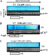
Detection of copper-binding proteins by MICS-BN-PAGE followed by copper ion detection PAGE. Each panel is divided into three parts. The top shows MICS-BN-PAGE (BN). The middle shows copper detection by PAGE for the whole BN lane; the probe complexed to copper is indicated (Cu2+), while the two upper signals correspond to Ca probe and free probe (probe). At the bottom is the Western blot (WB) of electroeluted protein from the MICS-BN-PAGE for CopI detection. (A) WT grown in 1.6 µM CuSO4; (B) WT in medium supplemented with 1.2 mM CuSO4; (C) the ΔcopI strain grown in 200 µM CuSO4. In panels A and C, no Cu2+-probe complex or CopI could be detected, while in panel B, in which CopI is induced, CopI and the Cu2+-probe complex are detected and colocalize in the same fractions.
FIG 4
FIG 4
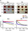
Growth phenotype and cell viability of the WT, ΔcopI mutant, and copA mutant grown with increasing copper concentrations. (A) Cells were grown in the dark with aerobic respiration (Res) (1.6 to 300 µM CuSO4) or anaerobically by PS (1.6 to 80 µM CuSO4) on plates for 48 h at 28°C prior to photography. (B) Copper tolerance was assayed for the WT, copA, and ΔcopI strains. Cells (initial optical density at 680 nm [OD680], 0.02) were grown in liquid by Res or by PS in the presence of different CuSO4 concentrations, and absorbance at 680 nm was measured after 24 h.
FIG 5
FIG 5
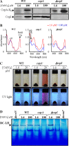
(A) Expression level of CopA and CopI in different genetic backgrounds. Equal amounts of proteins from WT CopAH6, ΔcopI CopAH6, and copA mutants were analyzed by Western blotting using the HRP-HisProbe. (B) Absorption spectra of total pigment extracts from WT, ΔcopI, and copA cells grown in malate or with 100 µM CuSO4. Pigments were extracted from the same amount of cells (1 OD680 unit). (C) Copper-dependent release of coproporphyrin III in the culture medium of grown cells. Phenotype of the ΔcopI mutant in comparison with the WT and the copA mutant grown in liquid malate or malate supplemented with 20 µM CuSO4. Cultures were centrifuged to reveal the presence of the UV-fluorescent pigment identified previously as coproporphyrin III (25) only in the spent medium of the copA mutant grown in the presence of 20 µM CuSO4. (D) Comparison of the amount of photosystem (RC-LH) in the solubilized membrane fractions of the WT, ΔcopI, and copA strains. Equal amounts of DDM-solubilized membranes were separated on 3-to-12% gradient BN-PAGE.
FIG 6
FIG 6
(A) cbb3 oxidase in-gel activity assay on BN-PAGE gradient. DAB staining was used to detect the cbb3 oxidase activity. (B) Detection of cbb3 subunits by Western blotting. The cbb3 CcoN, CcoO, and CcoP subunits were revealed in these membranes using specific antibodies raised against these subunits. (C) Detection of c-type cytochromes. The c-type cytochromes were revealed in the presence of TMBZ. For both panels B And C, equal amounts of membrane proteins from the WT and mutants were separated on 12% SDS–PAGE.
FIG 7
FIG 7
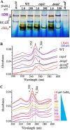
(A) Succinate dehydrogenase (SDH) in-gel activity assay. The membranes on BN-PAGE assayed for cbb3 oxidase (Fig. 6A) were subsequently assayed for SDH activity. (B) Difference (reduced minus oxidized) spectra of WT, ΔcopI, and copA membranes from cells grown in 1.6 µM (red) or 100 µM (blue) CuSO4. ΔccoN and ΔccmF strains were grown only in 1.6 µM CuSO4. (C) Difference spectra of total fractions from WT grown in the presence of increasing copper concentrations.
FIG 8
FIG 8
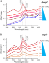
Difference in total b- and c-type cytochrome content between ΔcopI and copA mutants. Difference (reduced minus oxidized) spectra of total fractions from ΔcopI (A) and copA (B) grown microaerobically in the presence of increasing copper concentrations are shown.

References

    1. Macomber L, Imlay JA. 2009. The iron-sulfur clusters of dehydratases are primary intracellular targets of copper toxicity. Proc Natl Acad Sci U S A 106:8344–8349. doi:10.1073/pnas.0812808106. - DOI - PMC - PubMed
    1. Tan G, Cheng Z, Pang Y, Landry AP, Li J, Lu J, Ding H. 2014. Copper binding in IscA inhibits iron-sulphur cluster assembly in Escherichia coli. Mol Microbiol 93:629–644. doi:10.1111/mmi.12676. - DOI - PMC - PubMed
    1. Nies DH, Herzberg M. 2013. A fresh view of the cell biology of copper in enterobacteria. Mol Microbiol 87:447–454. doi:10.1111/mmi.12123. - DOI - PubMed
    1. González-Guerrero M, Raimunda D, Cheng X, Argüello JM. 2010. Distinct functional roles of homologous Cu+ efflux ATPases in Pseudomonas aeruginosa. Mol Microbiol 78:1246–1258. doi:10.1111/j.1365-2958.2010.07402.x. - DOI - PubMed
    1. Hassani BK, Astier C, Nitschke W, Ouchane S. 2010. CtpA, a copper-translocating P-type ATPase involved in the biogenesis of multiple copper-requiring enzymes. J Biol Chem 285:19330–19337. doi:10.1074/jbc.M110.116020. - DOI - PMC - PubMed

MeSH terms

Associated data

LinkOut - more resources