. 2015 Sep 22;6(5):e01491-15.
doi: 10.1128/mBio.01491-15.

Virome Capture Sequencing Enables Sensitive Viral Diagnosis and Comprehensive Virome Analysis

Affiliations

Virome Capture Sequencing Enables Sensitive Viral Diagnosis and Comprehensive Virome Analysis

Thomas Briese et al. mBio. .

Erratum in

Abstract

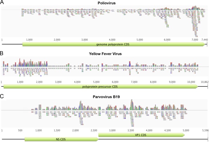
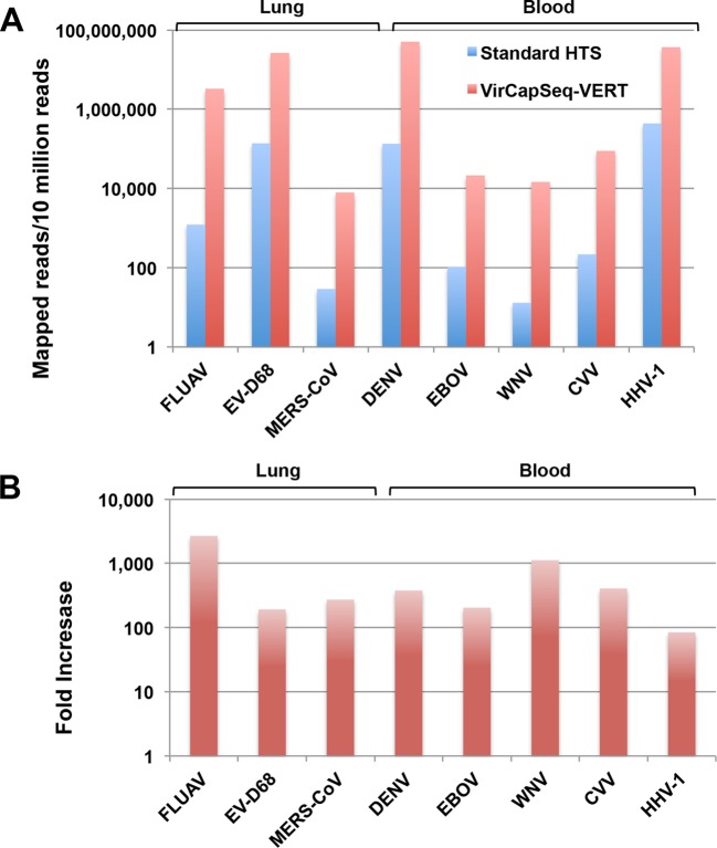
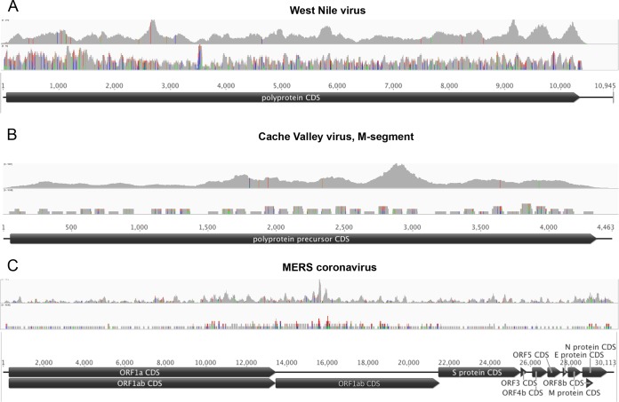
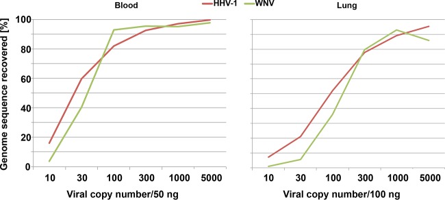
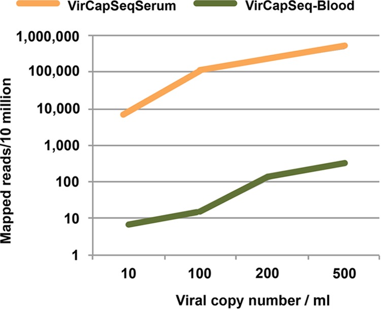
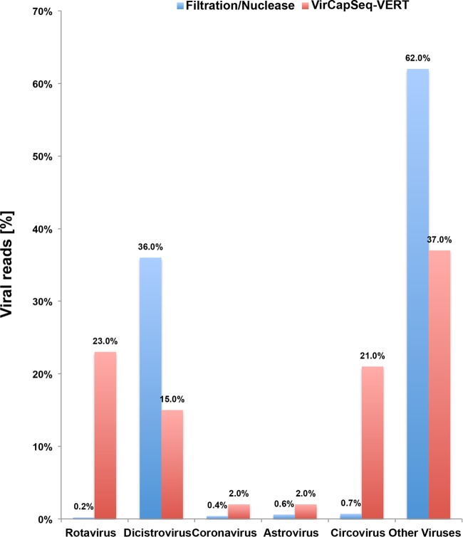
Insensitivity and technical complexity have impeded the implementation of high-throughput nucleic acid sequencing in differential diagnosis of viral infections in clinical laboratories. Here, we describe the development of a virome capture sequencing platform for vertebrate viruses (VirCapSeq-VERT) that increases the sensitivity of sequence-based virus detection and characterization. The system uses ~2 million probes that cover the genomes of members of the 207 viral taxa known to infect vertebrates, including humans. A biotinylated oligonucleotide library was synthesized on the NimbleGen cleavable array platform and used for solution-based capture of viral nucleic acids present in complex samples containing variable proportions of viral and host nucleic acids. The use of VirCapSeq-VERT resulted in a 100- to 10,000-fold increase in viral reads from blood and tissue homogenates compared to conventional Illumina sequencing using established virus enrichment procedures, including filtration, nuclease treatments, and RiboZero rRNA subtraction. VirCapSeq-VERT had a limit of detection comparable to that of agent-specific real-time PCR in serum, blood, and tissue extracts. Furthermore, the method identified novel viruses whose genomes were approximately 40% different from the known virus genomes used for designing the probe library. The VirCapSeq-VERT platform is ideally suited for analyses of virome composition and dynamics. IMPORTANCE : VirCapSeq-VERT enables detection of viral sequences in complex sample backgrounds, including those found in clinical specimens, such as serum, blood, and tissue. The highly multiplexed nature of the system allows both the simultaneous identification and the comprehensive genetic characterization of all known vertebrate viruses, their genetic variants, and novel viruses. The operational simplicity and efficiency of the VirCapSeq-VERT platform may facilitate transition of high-throughput sequencing to clinical diagnostic as well as research applications.


Figures

FIG 1
FIG 1
In silico validation of the VirCapSeq-VERT probe design. Probe depth and coverage of the VirCapSeq-VERT probe library are shown for poliovirus (A), yellow fever virus (B), and parvovirus B19 (C). Virus genomes are represented by black lines. The coding sequences are represented by green boxes. The probes are indicated by grey boxes. The top graph in each panel indicates probe depth at each locus. Colored lines in the probes indicate mismatch to the reference used for alignment (green, A; red, T; blue, C; orange, G). Line heights in the coverage track above indicate frequency of the mismatched bases.
FIG 2
FIG 2
VirCapSeq-VERT enhances the performance of high-throughput sequencing by increasing the number of mapped viral reads recovered from high-background specimens. Eight different viral NAs were quantitated by qPCR and used to spike a background of lung-derived (3 viruses) or blood-derived (5 viruses) NA extracts. Samples were split in two and processed by standard HTS (blue) or with VirCapSeq-VERT (red). FLUAV, influenza A virus; EVD-68, enterovirus D68; MERS-CoV, MERS coronavirus; DENV, dengue virus; EBOV, Ebola virus; WNV, West Nile virus; CVV, Cache Valley virus; HHV-1, human herpesvirus 1.
FIG 3
FIG 3
Read coverage versus probe coverage of VirCapSeq-VERT for West Nile virus (A), Cache Valley virus (B), and MERS coronavirus (C). Virus genomes are represented by horizontal black lines and coding sequence by black pointed boxes. The top graph in each panel indicates the read coverage obtained by VirCapSeq-VERT; probe coverage is shown below. Colored lines indicate mismatch to the reference used for alignment (green, A; red, T; blue, C; orange, G). Line heights indicate the frequency of the mismatched bases.
FIG 4
FIG 4
Limit of detection for VirCapSeq-VERT. Total nucleic acid from blood or lung tissue was spiked with human herpesvirus 1 (HHV-1) and West Nile virus (WNV) nucleic acid. The two preparations were serially diluted to generate six samples containing both viruses at 5,000, 1,000, 300, 100, 30, or 10 copies in 100 ng lung tissue or 50 ng whole-blood nucleic acid. Samples were processed with VirCapSeq-VERT.
FIG 5
FIG 5
Efficiency of viral read mapping with VirCapSeq-VERT. Human blood and serum were spiked with live enterovirus D68 virus stock quantitated by qPCR to generate samples with 500, 200, 100, or 10 copies/ml. Five hundred microliters of each sample was extracted and processed with VirCapSeq-VERT.
FIG 6
FIG 6
Selective enhancement of vertebrate virus detection by VirCapSeq-VERT. Bat fecal sample material was divided in two and analyzed using HTS with filtration and nuclease digest combined with postextraction DNase treatment or using VirCapSeq-VERT alone. VirCapSeq-VERT reduced the number of nonvertebrate viral reads and efficiently sequenced vertebrate virus sequences detected only at low levels by conventional HTS.

References

    1. Kapoor A, Lipkin WI. 2001. Virus discovery in the 21st century. In eLS. John Wiley & Sons, New York, NY.
    1. Mullis KB, Faloona FA. 1987. Specific synthesis of DNA in vitro via a polymerase-catalyzed chain reaction. Methods Enzymol 155:335–350. - PubMed
    1. VanDevanter DR, Warrener P, Bennett L, Schultz ER, Coulter S, Garber RL, Rose TM. 1996. Detection and analysis of diverse herpesviral species by consensus primer PCR. J Clin Microbiol 34:1666–1671. - PMC - PubMed
    1. Casas I, Tenorio A, Echevarría JM, Klapper PE, Cleator GM. 1997. Detection of enteroviral RNA and specific DNA of herpesviruses by multiplex genome amplification. J Virol Methods 66:39–50. doi:10.1016/S0166-0934(97)00035-9. - DOI - PubMed
    1. Liljas L, Tate J, Lin T, Christian P, Johnson JE. 2002. Evolutionary and taxonomic implications of conserved structural motifs between picornaviruses and insect picorna-like viruses. Arch Virol 147:59–84. doi:10.1007/s705-002-8303-1. - DOI - PubMed

Publication types

Substances