. 2015 Sep 14:8:2577-87.
doi: 10.2147/OTT.S82225. eCollection 2015.

Protein tyrosine phosphatase SHP-1 sensitizes EGFR/HER-2 positive breast cancer cells to trastuzumab through modulating phosphorylation of EGFR and HER-2

Affiliations

Protein tyrosine phosphatase SHP-1 sensitizes EGFR/HER-2 positive breast cancer cells to trastuzumab through modulating phosphorylation of EGFR and HER-2

Yifen Wu et al. Onco Targets Ther. .

Abstract

Background: Trastuzumab resistance in HER-2 positive breast cancer cells is closely related to overexpression of both epidermal growth factor receptor (EGFR) and human epidermal receptor (HER-2). SHP-1 has been demonstrated to downregulate tyrosine kinase activity including EGFR via its phosphatase function, but its effect on HER-2 activity is still unknown. Here, we examined the hypothesis that SHP-1 enhances the anticancer efficacy of trastuzumab in EGFR/HER-2 positive breast cancer cells through combining dual inhibition of EGFR and HER-2.

Methods: Trastuzumab-resistant breast cancer SKBr-3 cells were generated by long-term in vitro culture of SKBr-3cells in the presence of trastuzumab. The SHP-1 was ectopically expressed by stable transfection. The activity and expression of EGFR, HER-2, and downstream signaling pathways were tested by Western blot. Cell viability was examined by the 3-(4,5-dimethylthiazol-2-yl)-2,5-diphenyltetrazolium bromide (MTT) assay, and apoptosis was examined by flow cytometry. The binding between SHP-1 and EGFR/HER-2 was evaluated by immunoprecipitation assay and bimolecular fluorescence complementation. The effects of SHP-1 on tumorigenicity and trastuzumab sensitivity were confirmed via in vivo xenograft model.

Results: Trastuzumab-resistant SKBr-3 cells showed aberrant co-expression of EGFR and HER-2. Introduction of wild-type SHP-1 inhibited cell proliferation, clone formation, and promoted the apoptosis induced by trastuzumab. Meanwhile, SHP-1 overexpression reduced phosphorylation levels of EGFR and HER-2 both in parental and trastuzumab-resistant SKBr-3 cells. In vivo study showed an increased antitumor effect of trastuzumab in SHP-1 overexpressed xenografts. At last, we discovered that SHP-1 can make complexes with both EGFR and HER-2, and both phospho-EGFR and phosphor-HER-2 levels in wild-type SHP-1 immunoprecipitates were less than those in phosphatase-inactive SHP-1 (C453S) immunoprecipitates, indicating that EGFR and HER-2 are potential substrates of SHP-1.

Conclusion: Taken together, we have demonstrated that the SHP-1 is a negative regulatory factor of the tyrosine kinase activity of HER-2 and EGFR through inhibiting phosphorylation. Dual targeting of EGFR and HER-2, by combining trastuzumab with SHP-1 overexpression, may improve response in HER-2 overexpressing breast cancer cells that also express high levels of EGFR.

Keywords: EGFR; HER-2; SHP-1; breast cancer; drug resistance; trastuzumab.


Figures

Figure 1
Figure 1
Trastuzumab-resistant breast cancer SKBr-3 cells display increased EGFR and HER-2 expression and enhanced phosphorylation levels in response to trastuzumab. Notes: (A) MTT assays of parental SKBr-3 cells and the trastuzumab-resistant cells after treatment with increasing concentrations of trastuzumab. (B) Plate colony formation assays using parental and resistant cells in the presence of trastuzumab (10 μg/mL). (C) Confirmation of the differential expression of the indicated mRNA by qRT-PCR. Each mRNA was normalized to GAPDH. (D) Parental and resistant SKBr3 cells were treated for 24 hours with 5 μg/m trastuzumab. After lysis, protein levels were assessed using Western blotting techniques. (E) Quantification and comparing the gray value of Western blot results between parental group and trastuzumab-resistant group. The gray value of each protein was normalized to that of GAPDH, and the GV of each phosphorylated protein was normalized to the GV of its own corresponding total protein before comparing; the gray value of proteins in parental group were set as 1. Error bars represent the mean ± SD of triplicate experiments. Statistical significance was calculated using the Student’s t-test or ANOVA tests. ***P<0.001. Abbreviations: EGFR, epidermal growth factor receptor; HER-2, human epidermal receptor; MTT, 3-(4,5-dimethylthiazol-2-yl)-2,5-diphenyltetrazolium bromide; qRT-PCR, quantitative reverse transcription polymerase chain reaction; GAPDH, glyceraldehyde 3-phosphate dehydrogenase; GV, gray value; SD, standard deviation; ANOVA, analysis of variance; OD, optical density.
Figure 2
Figure 2
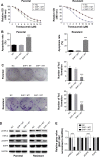
Overexpression of SHP-1 partly restores trastuzumab sensitivity in vivo. Notes: (A) MTT assays of modified SKBr-3 cells after treatment with increasing concentrations of trastuzumab. (B) Parental and resistant cells were treated with 5 μg/mL trastuzumab for 24 hours. Apoptosis rate in response was evaluated by using flow cytometry assay. (C) Plate colony formation assays of modified parental and trastuzumab-resistant cells were performed in the presence of 5 μg/mL trastuzumab. (D) Parental and resistant SKBr-3 cells were treated with 5 μg/mL trastuzumab, and then the total or phosphorylated EGFR and HER-2 protein levels were determined by Western blot. (E) Quantification and comparing the gray value of Western blot results between parental group and trastuzumab-resistant group. The gray value of each protein was normalized to that of GAPDH, and the GV of each phosphorylated protein was normalized to the GV of its own corresponding total protein before comparing, the gray value of proteins in NC group were set as 1. Error bars represent the mean ± SD of triplicate experiments. Statistical significance was calculated using the Student’s t-test or ANOVA tests. ***P<0.001. Abbreviations: MTT, 3-(4,5-dimethylthiazol-2-yl)-2,5-diphenyltetrazolium bromide; EGFR, epidermal growth factor receptor; HER-2, human epidermal receptor; GAPDH, glyceraldehyde 3-phosphate dehydrogenase; GV, gray value; NC, nonsense control; SD, standard deviation; ANOVA, analysis of variance; OD, optical density; WT, wild-type; MT, mutant-type.
Figure 3
Figure 3
SHP-1 binds to EGFR and HER-2 protein. Notes: (A, B) SKBR3 cells were transfected with control (NC), SHP-1 WT, or SHP-1 MT vector for 48 hours. EGFR and HER-2 proteins were pulled down by immunoprecipitation and the protein level was detected by immunoblotting. (C, D) SKBR3 cells were transfected with control (NC), SHP-1 WT, or SHP-1 MT vector for 48 hours. Phosphorylated-EGFR (pEGFR) and phosphorylated-HER-2 (pHER-2) proteins were pulled down by immunoprecipitation and the protein level was detected by immunoblotting. (E, F) The binding between SHP-1 and HER-3 or HER-4 was also evaluated through immunoprecipitation. The whole gel is shown, and no specific band of HER-3 or HER-4 was detected. (G) Bimolecular fluorescence complementation (BiFC) was performed to confirm the binding activity between SHP-1 and EGFR, HER-2, HER-3, and HER-4. BiFC signals were generated by interaction of the GFP fluorophore components based on proximity. + and − indicate receiving and withholding of the treatment, respectively. Abbreviations: EGFR, epidermal growth factor receptor; HER-2, human epidermal receptor; NC, nonsense control; WT, wild-type; IP, immunoprecipitation.
Figure 4
Figure 4
SHP-1 promotes trastuzumab sensitivity in vivo. Notes: Nude mice were inoculated in the mammary fat pad with trastuzumab-resistant SKBr-3 cells overexpressing SHP-1 or control scramble shRNA to allow tumor development. Mice were intravenously injected with 10 mg/kg trastuzumab twice a week. (A) One cohort of mice consisting five individuals in each treatment group were sacrificed after 45 days after first trastuzumab injection. (B) Tumor volume was recorded every 3 days, and (C) tumors were weighted and compared at last. (D) For survival assay, the survival of the other cohort containing ten mice in each group was recorded. Kaplan–Meier survival curves of the trastuzumab-treated mice are shown. ***P<0.001. Abbreviations: shRNA, small hairpin RNA; NC, nonsense control.

References

    1. Nahleh ZA. Hormonal therapy for male breast cancer: a different approach for a different disease. Cancer Treat Rev. 2006;32:101–105. - PubMed
    1. Malvezzi M, Bertuccio P, Levi F, La Vecchia C, Negri E. European cancer mortality predictions for the year 2013. Ann Oncol. 2013;24:792–800. - PubMed
    1. Yarden Y, Sliwkowski MX. Untangling the ErbB signalling network. Nat Rev Mol Cell Biol. 2001;2:127–137. - PubMed
    1. Slamon DJ, Godolphin W, Jones LA, et al. Studies of the HER-2/neu proto-oncogene in human breast and ovarian cancer. Science. 1989;244:707–712. - PubMed
    1. Slamon DJ, Clark GM, Wong SG, Levin WJ, Ullrich A, McGuire WL. Human breast cancer: correlation of relapse and survival with amplification of the HER-2/neu oncogene. Science. 1987;235:177–182. - PubMed