. 2015 Sep 11;9(5):054106.
doi: 10.1063/1.4930865. eCollection 2015 Sep.

Microfluidic point-of-care blood panel based on a novel technique: Reversible electroosmotic flow

Affiliations

Microfluidic point-of-care blood panel based on a novel technique: Reversible electroosmotic flow

Mahdi Mohammadi et al. Biomicrofluidics. .

Abstract

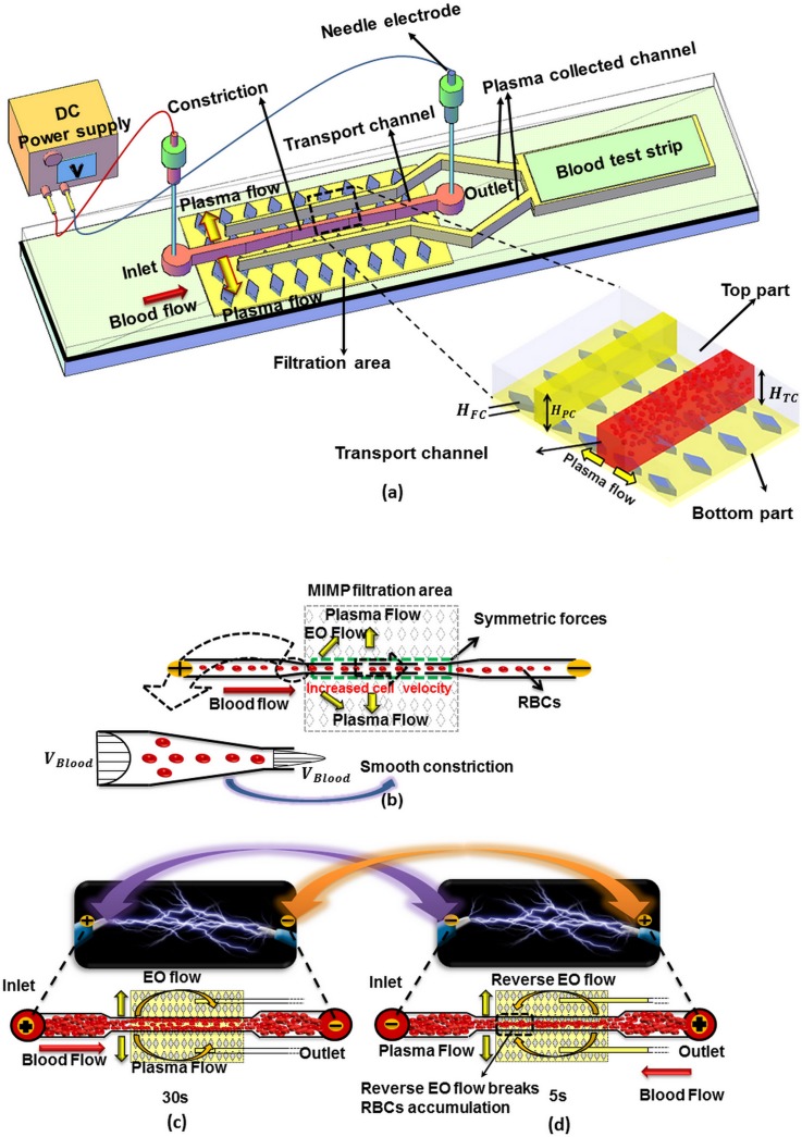
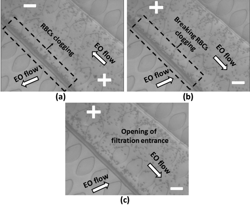
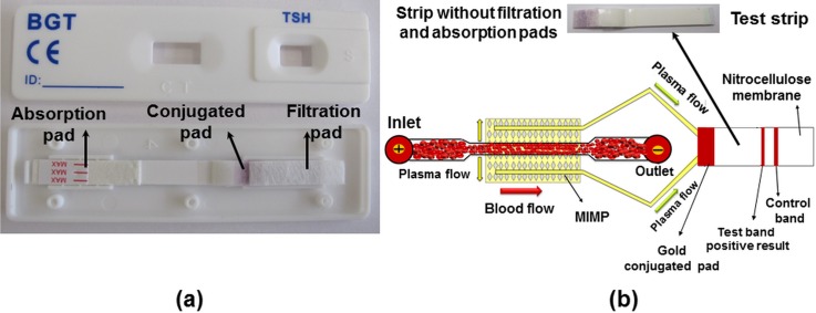
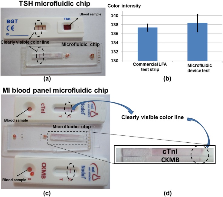
A wide range of diseases and conditions are monitored or diagnosed from blood plasma, but the ability to analyze a whole blood sample with the requirements for a point-of-care device, such as robustness, user-friendliness, and simple handling, remains unmet. Microfluidics technology offers the possibility not only to work fresh thumb-pricked whole blood but also to maximize the amount of the obtained plasma from the initial sample and therefore the possibility to implement multiple tests in a single cartridge. The microfluidic design presented in this paper is a combination of cross-flow filtration with a reversible electroosmotic flow that prevents clogging at the filter entrance and maximizes the amount of separated plasma. The main advantage of this design is its efficiency, since from a small amount of sample (a single droplet [Formula: see text]10 μl) almost 10% of this (approx 1 μl) is extracted and collected with high purity (more than 99%) in a reasonable time (5-8 min). To validate the quality and quantity of the separated plasma and to show its potential as a clinical tool, the microfluidic chip has been combined with lateral flow immunochromatography technology to perform a qualitative detection of the thyroid-stimulating hormone and a blood panel for measuring cardiac Troponin and Creatine Kinase MB. The results from the microfluidic system are comparable to previous commercial lateral flow assays that required more sample for implementing fewer tests.


Figures

FIG. 1.
FIG. 1.
(a) Schematic diagram of the blood plasma separation microdevice. (b) Detail of the transport channel constriction. (c) and (d) Electroosmotic flow and RBCs flow directions depending on the electrode polarization.
FIG. 2.
FIG. 2.
Design dimensions. (a) Top part. (b) Down part diamond shaped post-array.
FIG. 3.
FIG. 3.
Schematic view of the microdevice fabrication. (a)–(d) MIMP filtration channel manufacturing steps, (e) the microdevice top part with the test window, the channel inlet, and the channel outlet (PDMS), (f) the microdevice down part (glass), (g) bonding of both parts, and (h) the complete microdevice with the test strip covered with tape.
FIG. 4.
FIG. 4.
Microdevice performance. (a) Initial empty channel, (b) filling the transport channel after applying voltage and plasma separation, (c) filling the plasma-collected channel, and (d) end of the plasma-collected channel. (Multimedia view) [URL: http://dx.doi.org/10.1063/1.4930865.1]
FIG. 5.
FIG. 5.
RBC clogging and breakage procedure. (a) The RBCs clog the entrance of the filtration area. (b) Opening of the entrance of the filtration area using reverse EO flow. (c) The entrance is opened after 5 s. (Multimedia view) [URL: http://dx.doi.org/10.1063/1.4930865.2]
FIG. 6.
FIG. 6.
(a) All components of TSH test strip and (b) the test strip without filtration pad and the schematic view of microdevice with the test strip.
FIG. 7.
FIG. 7.
(a) The results of the TSH test using in the microfluidic chip and LFA strip. (b) The color intensity of the results of the TSH test using the microfluidic chip and the LFA strip. (c) and (d) The results of the myocardial infarction tests using the microfluidic chip and cTnI and CKMB LFA strips.

References

    1. Kersaudy-Kerhoas M. and Sollier E., Lab Chip 13(17), 3323–3346 (2013).10.1039/c3lc50432h - DOI - PubMed
    1. Gorkin R., Park J., Siegrist J., Amasia M., Lee B. S., Park J. M., Kim J., Kim H., Madou M., and Cho Y. K., Lab Chip 10(14), 1758–1773 (2010).10.1039/b924109d - DOI - PubMed
    1. Ducrée J., Haeberle S., Lutz S., Pausch S., Stetten F. V., and Zengerle R., J. Micromech. Microeng. 17, S103–S115 (2007).10.1088/0960-1317/17/7/S07 - DOI
    1. Haeberle S., Brenner T., Zengerle R., and Ducrée J., Lab Chip 6, 776–781 (2006).10.1039/b604145k - DOI - PubMed
    1. Vella S. J., Beattie P., Cademartiri R., Laromaine A., Martinez A. W., Phillips S. T., Mirica K., and Whitesides G. M., Anal. Chem. 84(6), 2883–2891 (2012).10.1021/ac203434x - DOI - PMC - PubMed

LinkOut - more resources