. 2015 Aug 15;7(8):1332-44.
eCollection 2015.

Tumstatin 185-191 increases the sensitivity of non-small cell lung carcinoma cells to cisplatin by blocking proliferation, promoting apoptosis and inhibiting Akt activation

Affiliations

Tumstatin 185-191 increases the sensitivity of non-small cell lung carcinoma cells to cisplatin by blocking proliferation, promoting apoptosis and inhibiting Akt activation

Wei Wang et al. Am J Transl Res. .

Abstract

Purpose: This study aimed to investigate the synergistic anti-tumor effects of tumstatin 185-191 and cisplatin in non-small cell lung carcinoma cells (NSCLC) (A549 cells and cisplatin resistant A549/DDP cells), and the potential role of Akt signaling pathway was also explored.

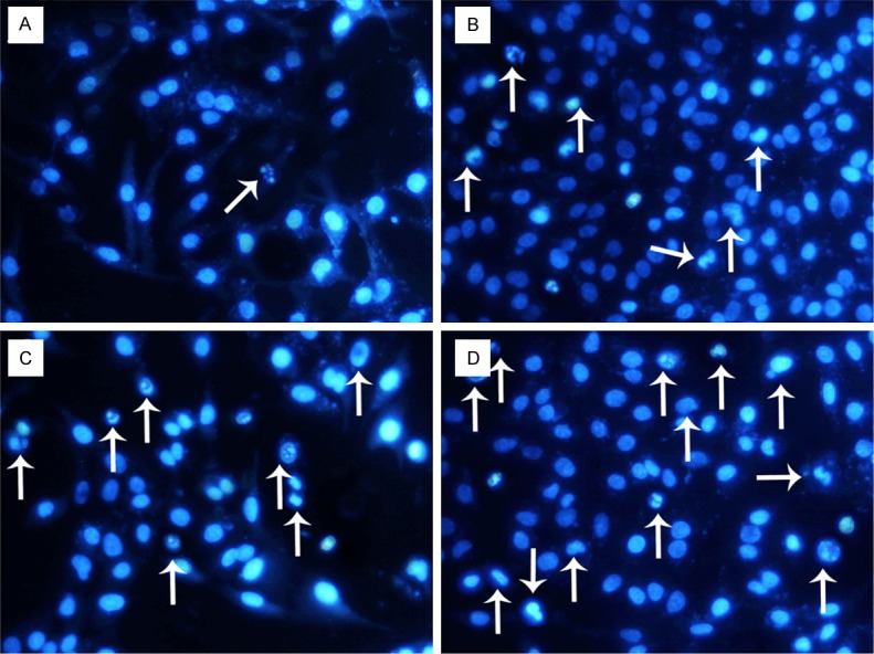
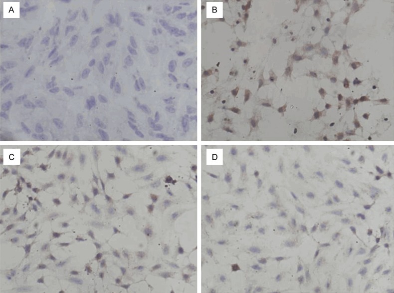
Methods: A549 or A549/DDP cells were treated with Tum185-191 or Tum185-191 plus cisplatin. Cell viability was assessed by modified MTT assay. 50% inhibiting concentration (IC50) and reversing drug-resistance index (RI) of chemotherapeutics were determined by MTT assay. Cell apoptosis was measured by Hoechst 33258 staining and flow cytometry. The activation of Akt signaling pathway was evaluated by immunocytochemistry and Western blot assay.

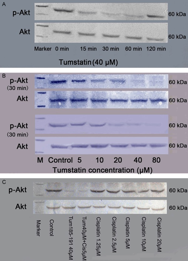
Results: Tum185-191 inhibited the proliferation of A549 cells and A549/DDP cells. In the presence of Tum185-191 (20 and 40 μM), IC50 of cisplatin reduced significantly in A549 cells and A549/DDP cells. Combined use of tumstatin 185-191 and cisplatin exerted synergistic effects in promoting apoptosis. A549 and A549/DDP cells had a high expression of p-Akt, and Tum185-191, but not cisplatin, significantly inhibited p-Akt expression. Combined use of cisplatin and Tum185-191 failed to further inhibit p-Akt expression. After Tum185-191 treatment, the increased p-Akt expression was observed at 15 min, peaked at 30-60 min, but disappeared at 120 min.

Conclusion: Tum185-191 increases the apoptosis, inhibit the proliferation, enhance the sensitivity of A549 cells to cisplatin and also partly reverse the resistance of A549-DDP cells to cisplatin, which is at least partially mediated by inactivating Akt pathway. These findings provide evidence for the chemotherapy of NSCLC with Tum185-191 and cisplatin.

Keywords: Akt; Non-small cell lung carcinoma cells; Tumstain 185-191; cisplatin.


Figures

Figure 1
Figure 1
A: Effects of Tum185-191 and cisplatin on the proliferation of A549 cells (48 h); B: Effects of Tum185-191 and cisplatin on the proliferation of A549/DDP cells (48 h).
Figure 2
Figure 2
Apoptosis of A549 cells treated with Tum185-191 and cisplatin for 48 h. (x 200, Arrow: apoptotic cells, Hoechst 33258 staining). A: Control group; B: 40 µM Tum185-191 group; C: 5 µM cisplatin; D: 40 µM Tums185-191 in combination with 5 µM cisplatin.
Figure 3
Figure 3
Apoptosis of A549/DDP cells treated with Tum185-191 and cisplatin for 48 h. (x 200, Arrow: apoptotic cells, Hoechst 33258 staining). A: Control group; B: 40 µM Tum185-191 group; C: 80 µM cisplatin; D: 40 µM Tums185-191 in combination with 80 µM cisplatin.
Figure 4
Figure 4
Apoptosis of A549 cells treated with Tum185-191 and cisplatin for 48 h (flow cytometry). A: Control group; B: 40 µM Tum185-191 group; C: 5 µM cisplatin; D: 40 µM Tums185-191 in combination with 5 µM cisplatin.
Figure 5
Figure 5
Apoptosis of A549/DDP cells treated with Tum185-191 and cisplatin for 48 h (flow cytometry). A: Control group; B: 40 µM Tum185-191 group; C: 80 µM cisplatin; D: 40 µM Tums185-191 in combination with 80 µM cisplatin.
Figure 6
Figure 6
p-Akt expression in A549 cells treated with Tum185-191 at different concentrations for 30 min (× 200, immunocytochemistry, p-Akt positive cellshaving brown granules). A: Blank control group; B: Control group; C: 20 µM Tum185-191 group; D: 40 µM Tums185-191 group.
Figure 7
Figure 7
p-Akt expression of in A549/DDP cells treated with Tum185-191 at different concentrations for 30 min (× 200, immunocytochemistry, positive cells having brown granules). A: Blank control group; B: Control group; C: 20 µM Tum185-191 group; D: 40 µM Tums185-191 group.
Figure 8
Figure 8
Western blot assay of p-Akt expression in A549 cells. A: A549 cells treated with 40 µM Tum 185-191; B: A549 cells treated with Tum185-191 for 30 and 60 min; C: A549 cells treated with Tum185-191 and cisplatin for 30 min.
Figure 9
Figure 9
Western blot assay of p-Akt expression in A549 cells. A: Untreated A549 cells and A549/DDP cells; B: A549/DDP cells treated with 40 µM Tum185-191; C: A549/DDP cells treated with Tum185-191 for 30 and 60 min; D: A549/DDP cells treated with Tum185-191 and cisplatin for 30 min.

References

    1. Ridge CA, McErlean AM, Ginsberg MS. Epidemiology of lung cancer. Semin Intervent Radiol. 2013;30:93–98. - PMC - PubMed
    1. Paliogiannis P, Attene F, Cossu A, Budroni M, Cesaraccio R, Tanda F, Trignano M, Palmieri G. Lung cancer epidemiology in North Sardinia, Italy. Multidiscip Respir Med. 2013;8:45. - PMC - PubMed
    1. Siegel R, Naishadham D, Jemal A. Cancer statistics, 2013. CA Cancer J Clin. 2013;63:11–30. - PubMed
    1. Dela Cruz CS, Tanoue LT, Matthay RA. Lung cancer: epidemiology, etiology, and prevention. Clin Chest Med. 2011;32:605–644. - PMC - PubMed
    1. Powell CA, Halmos B, Nana-Sinkam SP. Update in lung cancer and mesothelioma 2012. Am J Respir Crit Care Med. 2013;188:157–166. - PMC - PubMed

LinkOut - more resources