. 2015 Jul 15;5(8):2431-40.
eCollection 2015.

Novel 5-fluorouracil-resistant human esophageal squamous cell carcinoma cells with dihydropyrimidine dehydrogenase overexpression

Affiliations

Novel 5-fluorouracil-resistant human esophageal squamous cell carcinoma cells with dihydropyrimidine dehydrogenase overexpression

Osamu Kikuchi et al. Am J Cancer Res. .

Abstract

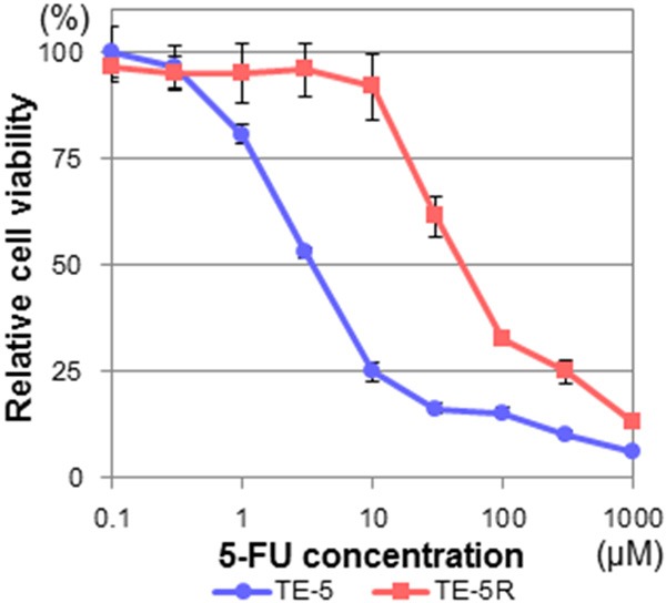
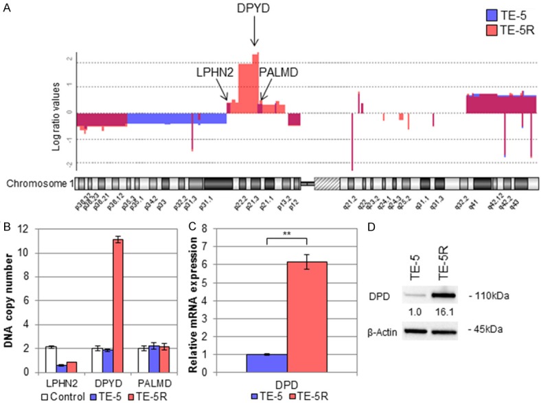
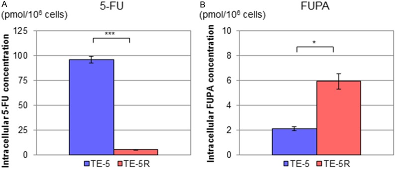
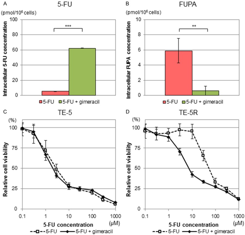
5-Fluorouracil (5-FU) is a key drug for the treatment of esophageal squamous cell carcinoma (ESCC); however, resistance to it remains a critical limitation to its clinical use. To clarify the mechanisms of 5-FU resistance of ESCC, we originally established 5-FU-resistant ESCC cells, TE-5R, by step-wise treatment with continuously increasing concentrations of 5-FU. The half maximal inhibitory concentration of 5-FU showed that TE-5R cells were 15.6-fold more resistant to 5-FU in comparison with parental TE-5 cells. TE-5R cells showed regional copy number amplification of chromosome 1p including the DPYD gene, as well as high mRNA and protein expressions of dihydropyrimidine dehydrogenase (DPD), an enzyme involved in 5-FU degradation. 5-FU treatment resulted in a significant decrease of the intracellular 5-FU concentration and increase of the concentration of α-fluoro-ureidopropionic acid (FUPA), a metabolite of 5-FU, in TE-5R compared with TE-5 cells in vitro. Conversely, gimeracil, a DPD inhibitor, markedly increased the intracellular 5-FU concentration, decreased the intracellular FUPA concentration, and attenuated 5-FU resistance of TE-5R cells. These results indicate that 5-FU resistance of TE-5R cells is due to the rapid degradation of 5-FU by DPD overexpression. The investigation of 5-FU-resistant ESCC with DPYD gene copy number amplification and consequent DPD overexpression may generate novel biological evidence to explore strategies against ESCC with 5-FU resistance.

Keywords: 5-fluorouracil; Esophageal squamous cell carcinoma; chemotherapy; dihydropyrimidine dehydrogenase; drug resistance.


Figures

Figure 1
Figure 1
5-FU resistance of TE-5R cells. TE-5 and TE-5R cells were treated with the indicated concentrations of 5-FU for 72 h, and cell viability was assessed using the WST-1 assay. A viability of 100% was defined as the amount of absorption at 450 nm in untreated cells. The mean value ± S.D. of six replicate wells from a representative experiment is shown. Each experiment was repeated at least three times, and consistent results were obtained. IC50 values of TE-5 and TE-5R cells were 3.6 ± 1.1 and 55.5 ± 10.1 μM, respectively. Note that TE-5R cells were 15.6-fold more resistant to 5-FU in comparison with parental TE-5 cells.
Figure 2
Figure 2
Amplification of DPYD gene and subsequent DPD expression in TE-5R cells. A. Chromosome 1 copy number alteration in aCGH analysis. The vertical axis indicates the log (base 2) ratio of DNA expression. TE-5R harbored specific regional amplification of the short arm of chromosome 1 around 1p2 including the DPYD gene (1p21.3). Note that LPHN2 and PALMD genes are not located in the specific amplified regions. B. qPCR analysis using LPHN2, DPYD and PALMD gene probes. The DPYD gene is amplified to produce 10 copies in DNA samples derived from TE-5R cells, but not non-cancerous human genomic DNA or parental TE-5 cells. On the other hand, LPHN2 and PALMD genes are not amplified in any samples. C. DPD mRNA expression levels in TE-5 and TE-5R cells. qPCR revealed significantly increased DPD mRNA expression in TE-5R compared with TE-5 cells. **P < 0.01. D. DPD protein expression levels in TE-5 and TE-5R cells. The relative density was calculated by densitometry. β-Actin served as a loading control. The DPD protein expression level in TE-5R cells was higher than in TE-5 cells.
Figure 3
Figure 3
Intracellular 5-FU and FUPA concentrations in TE-5 and TE-5R cells treated with 5-FU. TE-5 and TE-5R cells (5 × 10⁵ cells) were seeded in 6-well plates, and then treated with 5-FU (10 μM) for 24 h. Cells were harvested, and levels of 5-FU and the FUPA concentration were measured with liquid chromatography-tandem mass spectrometry. A. Intracellular 5-FU concentrations in TE-5 and TE-5R cells treated with 5-FU. Intracellular 5-FU concentrations in TE-5R cells were significantly lower than those in TE-5 cells (n = 3). ***P < 0.001. B. Intracellular FUPA concentrations in TE-5 and TE-5R cells treated with 5-FU. Intracellular FUPA concentrations in TE-5R cells were significantly higher than in TE-5 cells (n = 3). *P < 0.05.
Figure 4
Figure 4
Effects of DPD inhibitor on TE-5R cells. A DPD inhibitor, gimeracil, was added to the culture medium with the indicated concentrations of 5-FU at a molar ratio of 1:0.2 (5-FU:gimeracil) for 24 h. (i.e., 2 μM gimeracil to 10 μM 5-FU). (A) Intracellular 5-FU concentrations in TE-5R cells in the presence or absence of gimeracil. Intracellular 5-FU concentrations in TE-5R cells treated with 5-FU and gimeracil were markedly higher than those treated with 5-FU alone. (n = 3). ***P < 0.001. (B) Intracellular FUPA concentrations in TE-5R cells in the presence or absence of gimeracil. Intracellular FUPA concentrations in TE-5R cells treated with 5-FU and gimeracil were significantly lower than those treated with 5-FU alone. (n = 3). **P < 0.01. (C, D) Effects of gimeracil on 5-FU sensitivity of TE-5 (C) and TE-5R (D) cells. Gimeracil was added to the culture medium with the indicated concentrations of 5-FU at a molar ratio of 1:0.2 (5-FU:gimeracil) for 72 h. Cell viability was assessed by the WST-1 assay. A viability of 100% was defined as the amount of absorption at 450 nm in untreated cells. Each point represents the mean ± S.D. of sextuplicate wells. Note that gimeracil significantly attenuated 5-FU resistance in TE-5R but not TE-5 cells. Representative data from three independent experiments are shown.

Similar articles

Cited by

References

    1. Napier KJ, Scheerer M, Misra S. Esophageal cancer: A Review of epidemiology, pathogenesis, staging workup and treatment modalities. World J Gastrointest Oncol. 2014;6:112–120. - PMC - PubMed
    1. Pennathur A, Gibson MK, Jobe BA, Luketich JD. Oesophageal carcinoma. Lancet. 2013;381:400–412. - PubMed
    1. Enzinger PC, Mayer RJ. Esophageal cancer. N Engl J Med. 2003;349:2241–2252. - PubMed
    1. Pennathur A, Farkas A, Krasinskas AM, Ferson PF, Gooding WE, Gibson MK, Schuchert MJ, Landreneau RJ, Luketich JD. Esophagectomy for T1 esophageal cancer: outcomes in 100 patients and implications for endoscopic therapy. Ann Thorac Surg. 2009;87:1048–1054. - PMC - PubMed
    1. Ancona E, Ruol A, Santi S, Merigliano S, Sileni VC, Koussis H, Zaninotto G, Bonavina L, Peracchia A. Only pathologic complete response to neoadjuvant chemotherapy improves significantly the long term survival of patients with resectable esophageal squamous cell carcinoma: final report of a randomized, controlled trial of preoperative chemotherapy versus surgery alone. Cancer. 2001;91:2165–2174. - PubMed