

Telocytes in the Spleen
- PMID: 26397114
- PMCID: PMC4580423
- DOI: 10.1371/journal.pone.0138851
Telocytes in the Spleen
Abstract
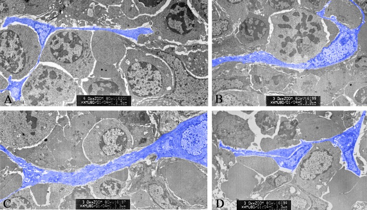
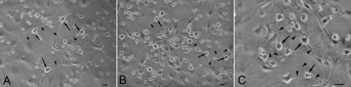
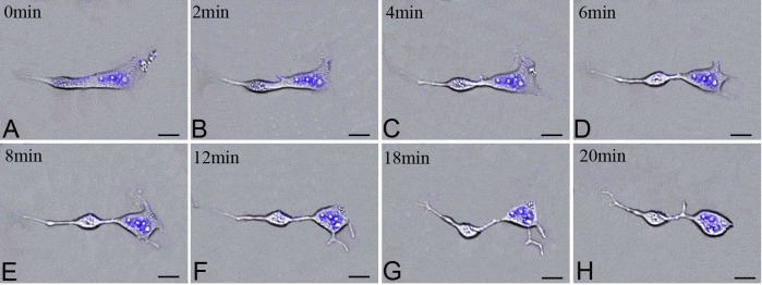
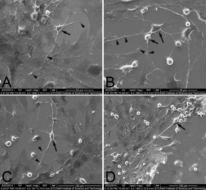
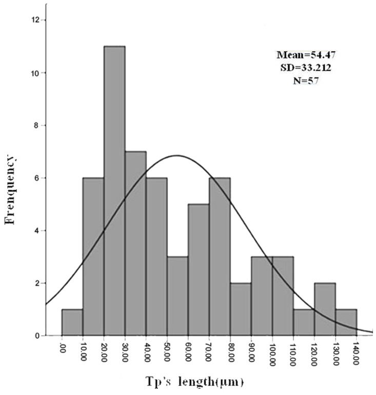
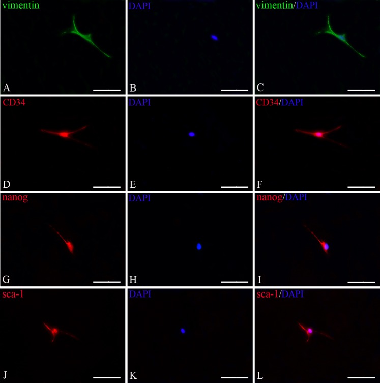
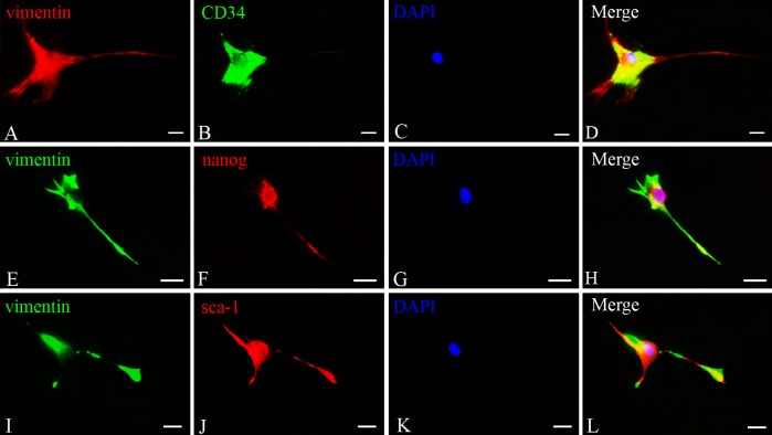
Telocytes, a novel type of interstitial cells with very long and thin prolongations, have been identified in many organs in mammals. At present, the ultrastructural, immunocytochemical and electrophysiological properties of telocytes in multiple organs have been understood. However, telocytes in spleen, especially their roles in spleen have not been reported. The aim of this study was to investigate the ultrastructure, distribution and immunophenotypes of splenic telocytes. Rat spleen was harvested for the ultrastructure analysis by transmission electron microscopy (TEM). The primary culture of telocytes was performed after combined enzymatic digestion. The characteristic morphology was analyzed by a scanning electron microscopy (SEM). It was shown that telocytes displayed a piriform/spindle/triangular shape with long and slender telopods and extremely long prolongation contracting with surrounding cells in the spleen. Their dynamic profiles of cytoplasmic separation were recorded by the Live Cell Imaging System. The length of telopods was mostly distributing in 20-30 μm, in accordance with normal distribution. Most telocytes had three or two telopods (28.71% and 22.58% respectively). Immunostaining indicated that these cells were positive for vimentin, CD34, nanog and sca-1, but negative for c-kit. These data prove the existence of telocytes in the spleen, which may serve as the experimental base for exploring their roles in the spleen.
Conflict of interest statement
Figures
Similar articles
-
Multiple immunophenotypes of cardiac telocytes.Exp Cell Res. 2015 Nov 1;338(2):239-44. doi: 10.1016/j.yexcr.2015.08.012. Epub 2015 Aug 21. Exp Cell Res. 2015. PMID: 26302265
-
Identification and protective role of CD34+ stromal cells/telocytes in experimental autoimmune encephalomyelitis (EAE) mouse spleen.Histochem Cell Biol. 2023 Jul;160(1):11-25. doi: 10.1007/s00418-023-02186-5. Epub 2023 Apr 4. Histochem Cell Biol. 2023. PMID: 37014442
-
CD34+ stromal cells/telocytes and their role in mouse lung development: Light microscopy, immunofluorescence, ultrastructural and scanning electron microscopy evidence.Cell Biol Int. 2024 Nov;48(11):1680-1697. doi: 10.1002/cbin.12223. Epub 2024 Aug 4. Cell Biol Int. 2024. PMID: 39099163
-
The Cutaneous Telocytes.Adv Exp Med Biol. 2016;913:303-323. doi: 10.1007/978-981-10-1061-3_20. Adv Exp Med Biol. 2016. PMID: 27796896 Review.
-
Features of Telocytes in Agricultural Animals.Adv Exp Med Biol. 2016;913:105-113. doi: 10.1007/978-981-10-1061-3_6. Adv Exp Med Biol. 2016. PMID: 27796882 Review.
Cited by
-
Expression of toll-like receptors and their regulatory roles in murine cardiac telocytes.J Cell Mol Med. 2019 Aug;23(8):5360-5368. doi: 10.1111/jcmm.14416. Epub 2019 Jun 24. J Cell Mol Med. 2019. PMID: 31232516 Free PMC article.
-
Immunohistochemical biomarkers and distribution of telocytes in ApoE-/- mice.Cell Biol Int. 2019 Nov;43(11):1286-1295. doi: 10.1002/cbin.11128. Epub 2019 Jun 28. Cell Biol Int. 2019. PMID: 30912221 Free PMC article.
-
Telocytes Enhances M1 Differentiation and Phagocytosis While Inhibits Mitochondria-Mediated Apoptosis Via Activation of NF-κB in Macrophages.Cell Transplant. 2021 Jan-Dec;30:9636897211002762. doi: 10.1177/09636897211002762. Cell Transplant. 2021. PMID: 33787355 Free PMC article.
-
Recently Discovered Interstitial Cell Population of Telocytes: Distinguishing Facts from Fiction Regarding Their Role in the Pathogenesis of Diverse Diseases Called "Telocytopathies".Medicina (Kaunas). 2019 Feb 18;55(2):56. doi: 10.3390/medicina55020056. Medicina (Kaunas). 2019. PMID: 30781716 Free PMC article. Review.
-
Telocytes: a potential defender in the spleen of Npc1 mutant mice.J Cell Mol Med. 2017 May;21(5):848-859. doi: 10.1111/jcmm.13024. Epub 2016 Nov 18. J Cell Mol Med. 2017. PMID: 27860245 Free PMC article.
References
Publication types
MeSH terms
Substances
LinkOut - more resources
Full Text Sources
Other Literature Sources
Research Materials
