Comparative Study
. 2015 Oct 13;6(31):31702-20.
doi: 10.18632/oncotarget.5165.

KDM4B and KDM4A promote endometrial cancer progression by regulating androgen receptor, c-myc, and p27kip1

Affiliations
Comparative Study

KDM4B and KDM4A promote endometrial cancer progression by regulating androgen receptor, c-myc, and p27kip1

Mei-Ting Qiu et al. Oncotarget. .

Abstract

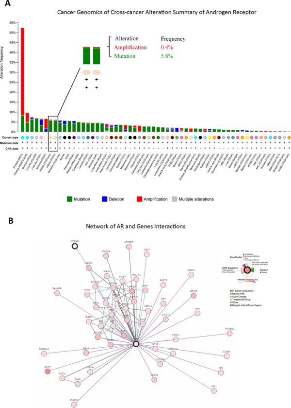
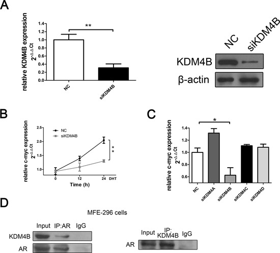
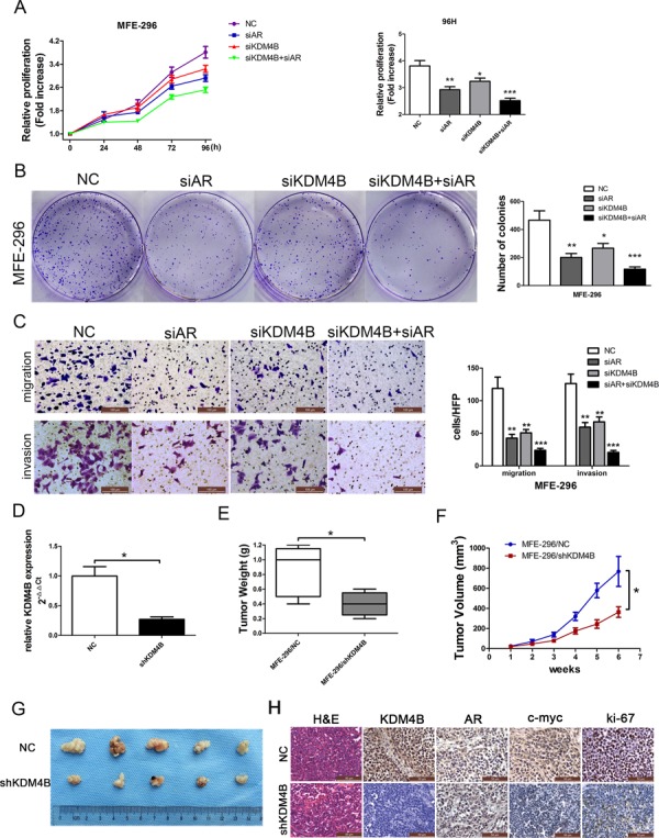
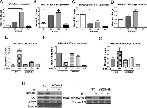
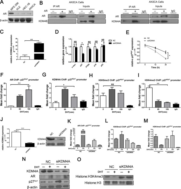
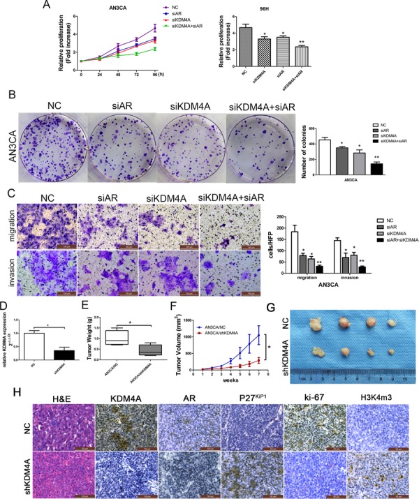
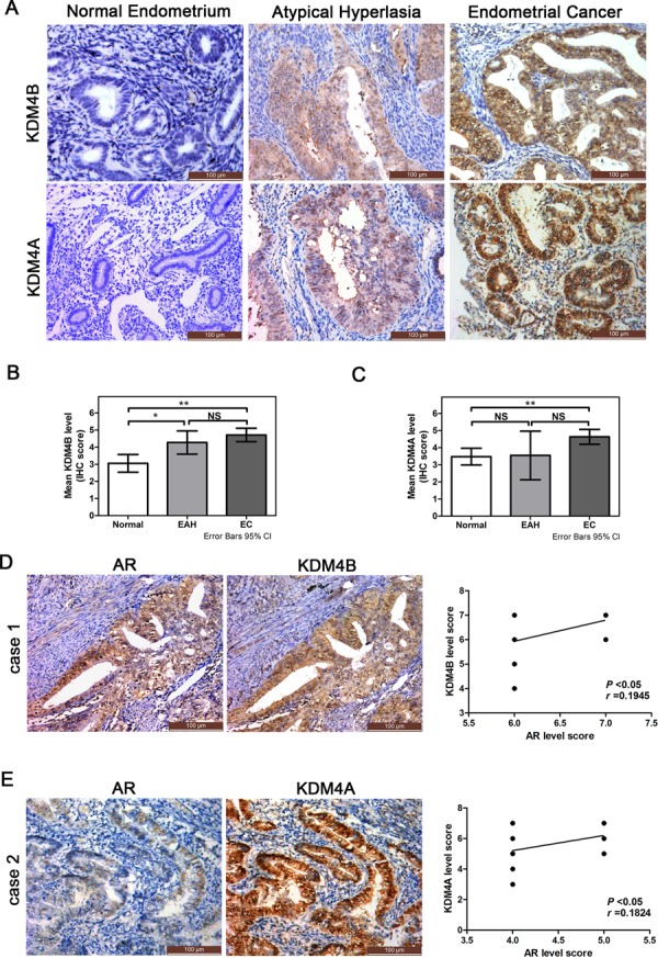
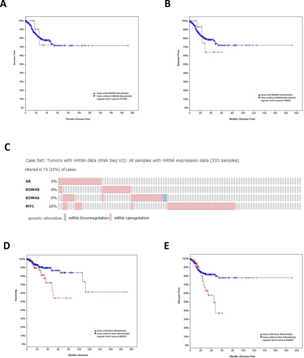
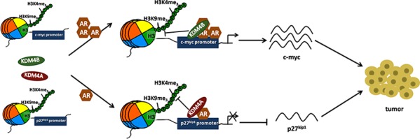
Epidemiological evidence suggests that elevated androgen levels and genetic variation related to the androgen receptor (AR) increase the risk of endometrial cancer (EC). However, the role of AR in EC is poorly understood. We report that two members of the histone demethylase KDM4 family act as major regulators of AR transcriptional activityin EC. In the MFE-296 cell line, KDM4B and AR upregulate c-myc expression, while in AN3CA cells KDM4A and AR downregulate p27kip1. Additionally, KDM4B expression is positively correlated with AR expression in EC cell lines with high baseline AR expression, while KDM4A and AR expression are positively correlated in low-AR cell lines. In clinical specimens, both KDM4B and KDM4A expression are significantly higher in EC tissues than that in normal endometrium. Finally, patients with alterations in AR, KDM4B, KDM4A, and c-myc have poor overall and disease-free survival rates. Together, these findings demonstrate that KDM4B and KDM4A promote EC progression by regulating AR activity.

Keywords: androgen receptor; c-myc; endometrial cancer; histone modification; lysine demethylases KDM4B and KDM4A; p27kip1; prognosis.


Conflict of interest statement

CONFLICTS OF INTEREST

The authors declare that they have no conflicts of interest.

Figures

Figure 1
Figure 1. In silico analysis of patient database identified novel AR-KDM4B signaling in EC
A. Genomics of cross-cancer alteration summary of AR in all cancers, which included AR amplification, mutation, and deletion. B. AR and relative gene interactions network using existing data from The Cancer Genome Atlas.
Figure 2
Figure 2. KDM4B binds with AR and activates AR-mediated transcription in MFE-296 EC cells
A. KDM4B knockdown was confirmed by qRT-PCR and Western blotting in MFE-296 cells. β-actin was used as a loading control. B. MFE-296 cells were transiently transfected with either negative control (NC) or KDM4B (siKDM4B) siRNA in steroid-depleted media and treated with 100 nM DHT for up to 24 h before RNA extraction. qRT-PCR was used to assess c-myc mRNA expression. C. KDM4B silencing inhibited c-myc mRNA expression in MFE-296 cells, whereas knockdown of other KDM4 enzymes didn't affect c-myc expression. D. MFE-296 cells grown in serum-containing media were subject to co-IP using anti-AR, anti-KDM4B, or control antibodies before Western blotting using reciprocal antibodies. All experiments were performed two or more times, and data represent the mean fold change ± SE.
Figure 3
Figure 3. KDM4B promotes AR-mediated carcinogenesis in MFE-296 cells
MFE-296 cells were transiently transfected with negative control (NC), AR (siAR) siRNA, KDM4B (siKDM4B) siRNA or KDM4B and AR siRNA together (siKDM4B+siAR). Cell proliferation was determined by MTT assay A. and colony formation assays B, C. Migrated and invasive MFE-296 cells on the lower surface of the Transwell filter were stained and photographed, 200×. The number of migrated and invasive cells is shown on the right. *P < 0.05, **P < 0.01, ***P < 0.005 compared with the NC group. D. KDM4B knockdown efficiency in shKDM4B group was confirmed by qRT-PCR. The tumor weight E. and tumor volumes F. and G. formed from nude mice injected subcutaneously with MFE-296 cells stably transfected with NC (MFE-296/NC) or shKDM4B (MFE-296/shKDM4B) were shown. H. Staining with hematoxylin and eosin (H&E) or immunohistochemical staining for KDM4B, AR, c-myc, and ki-67 in mouse tumor tissues (400×).
Figure 4
Figure 4. KDM4B activates AR target c-myc by demethylating H3K9me3 in response to androgens in MFE-296 cells
A series of ChIP assays for A. AR, B. KDM4B, C. H3K9me3, and D. H3K4me3 at the c-myc promoter in MFE-296 cells treated with DHT (100 nM) for 0, 60, or 180 min. *P < 0.05 compared with 0 min. MFE-296 cells were transiently transfected with either negative control (NC) or KDM4B siRNA (siKDM4B) and placed in steroid-depleted media for 72 h with or without a 180 min exposure to 100 nM DHT. ChIP was then performed with an anti-AR antibody E. an anti-H3K9me3 antibody F. or an anti-H3K4me3 antibody G. and analyzed at the c-myc promoter. Nonspecific IgG was used as a negative control. H. MFE-296 cells were subject to transient transfection with either negative control or KDM4B (siKDM4B) siRNAs in steroid-depleted media for 48 h before a 24 h to 100nM DHT. After protein extraction, AR, KDM4B, c-myc (H), and global histone modifications I. were assessed by Western blotting. Histone H3 and β-actin were used as loading controls.
Figure 5
Figure 5. KDM4A, but not KDM4B, binds with AR and suppresses AR target p27kip1 by demethylating H3K4me3 in response to androgens in AN3CA cells
A. AR expression in various EC cell lines; AN3CA cells were studied because of their low AR expression. B. AN3CA cells transiently transfected with mammalian expression vectors encoding AR or empty vector control were subject to co-IP using anti-AR or control antibodies before Western blotting using anti-KDM4B (B, left) or anti-KDM4A (B, right). C. KDM4A over-expression was confirmed by qRT-PCR in AN3CA cells. D. AN3CA cells were transiently transfected with either negative control (NC) or KDM4A expression plasmid (exKDM4A). Overexpression of KDM4A inhibited p27kip1 mRNA expression in qRT-PCR assays in AN3CA cells E. the expression of other suppressor genes, including c-myc, did not affect p27kip1 levels (D) *P < 0.05 compared with NC. ChIP for F. AR, G. KDM4A, H. H3K9me3, and I. H3K4me3 at the p27kip1 promoter in AN3CA cells treated with DHT (100 nM) for 0, 60, or 180 min. *P < 0.05, **P < 0.01 compared with 0 min. J. AN3CA cells were transiently transfected with either negative control (NC) or KDM4A siRNA (siKDM4A) in steroid-depleted media for 72 h with or without 180 min of exposure to 100 nM DHT before ChIP with an anti-AR antibody K. an anti-H3K9me3 antibody L. or an anti- H3K4me3 antibody M. and subsequently analyzed at the p27kip1 promoter. Nonspecific IgG was used as a negative control. N. AN3CA cells were subject to transient transfection with either negative control or KDM4A (siKDM4A) siRNAs in steroid-depleted media for 48 h before 24 h of exposure to 100nM DHT followed by protein extraction. AR, KDM4A, p27kip1 (N) and global histone modifications O. were assessed with Western blots. Histone H3 and β-actin were used as loading controls.
Figure 6
Figure 6. KDM4A, but not KDM4B, promotes AR-mediated carcinogenesis in AN3CA cells
A. AN3CA cells were transiently transfected with negative control (NC), AR (siAR) siRNA, KDM4A (siKDM4A) siRNA or KDM4A and AR siRNA together (siKDM4A+siAR). Cell proliferation was determined by the MTT assay (A) and colony formation assays B, C. Migrated and invasive AN3CA cells on the lower surface of the Transwell filter were stained and photographed, 200×. The number of migrated and invasive cells is shown on the right. *P < 0.05, **P < 0.01 compared with the NC group. D. KDM4A knockdown efficiency in shKDM4A group was confirmed by qRT-PCR. Tumor weight E. and tumor volumes F. and G. from nude mice injected subcutaneously with AN3CA cells stably transfected with NC (AN3CA/NC) or shKDM4A (AN3CA/shKDM4A). H. Staining with hematoxylin and eosin (H&E) or immunohistochemical staining for KDM4A, AR, p27kip1, ki-67 and H3k4me3 in mouse tumor tissues (400×).
Figure 7
Figure 7. Expression of KDM4B and KDM4A in endometrial tissues and their relationship to AR expression in EC specimens
A. Immunohistochemical stain of KDM4B and KDM4A in normal endometrium, endometrial atypical hyperplasia (EAH), and endometrioidcancer (EC) (200×). Summary of the immunostaining scores of B. KDM4B and C. KDM4A in normal endometrium, EAH and EC (*P < 0.05, **P < 0.01; NS, not significant). A score of ≥4 was considered positive for KDM4B and KDM4A expression. D. Example of KDM4B and AR immunoreactivity in EC tissues. KDM4B and AR levels were correlated in the 21 ECs with high AR scores (6 or 7) (r = 0.1945, P < 0.05). E. Example of KDM4A and AR immunoreactivity in EC tissues. KDM4A and AR levels were correlated in the 29 ECs with lower AR scores (4 or 5) (r = 0.1824, P < 0.05).
Figure 8
Figure 8. In silico analysis of OS and DFS in EC patients
The Kaplan-Meier disease-free survival (DFS) of uterine corpus endometrioid carcinoma patients (n = 333) from a dataset in The Cancer Genome Atlas stratified by A. KDM4B and B. KDM4A expression. C. OncoPrint of AR, KDM4B, KDM4A, c-myc pathway alterations in EC. Genomic alteration provides an overview of genomic alterations (legend) in particular genes (rows) affecting particular individual sample (columns). The Kaplan-Meier D. overall survival (OS) and E. DFS of uterine corpus endometrioid carcinoma patients from a dataset in The Cancer Genome Atlas stratified by AR, KDM4B, KDM4A, and c-myc alteration.
Figure 9
Figure 9. Schematic of KDM4B and KDM4A regulation of AR signaling in EC
In EC tissues with high levels of AR, KDM4B, along with AR, is recruited to cis-regulatory elements of AR-target gene c-myc and activates c-myc expression by H3K9me3 demethylation. In EC tissues with low level of AR, KDM4A, along with AR, is recruited to cis-regulatory elements of AR-target gene p27kip1 and decreases expression of this tumor suppressor by H3K4me3 demethylation. These alterations in c-myc and p27kip1 expression promote EC progression.

References

    1. Siegel RL, Miller KD, Jemal A. Cancer statistics, 2015. CA Cancer J Clin. 2015;65:5–29. - PubMed
    1. Whitney CW, Brunetto VL, Zaino RJ, Lentz SS, Sorosky J, Armstrong DK, Lee RB. Phase II study of medroxyprogesterone acetate plus tamoxifen in advanced endometrial carcinoma: a Gynecologic Oncology Group study. Gynecol Oncol. 2004;92:4–9. - PubMed
    1. Erkanli S, Ayhan A. Fertility-sparing therapy in young women with endometrial cancer: update. Int J Gynecol Cancer. 2010;20:1170–1187. - PubMed
    1. Fearnley EJ, Marquart L, Spurdle AB, Weinstein P, Webb PM. Polycystic ovary syndrome increases the risk of endometrial cancer in women aged less than 50 years: an Australian case-control study. Cancer causes & control: CCC. 2010;21:2303–2308. - PubMed
    1. Tuckerman EM, Okon MA, Li T, Laird SM. Do androgens have a direct effect on endometrial function? An in vitro study. Fertil Steril. 2000;74:771–779. - PubMed

Publication types

MeSH terms