. 2015 Oct 20;6(32):33397-409.
doi: 10.18632/oncotarget.5179.

BRD4-targeted therapy induces Myc-independent cytotoxicity in Gnaq/11-mutatant uveal melanoma cells

Affiliations

BRD4-targeted therapy induces Myc-independent cytotoxicity in Gnaq/11-mutatant uveal melanoma cells

Grazia Ambrosini et al. Oncotarget. .

Abstract

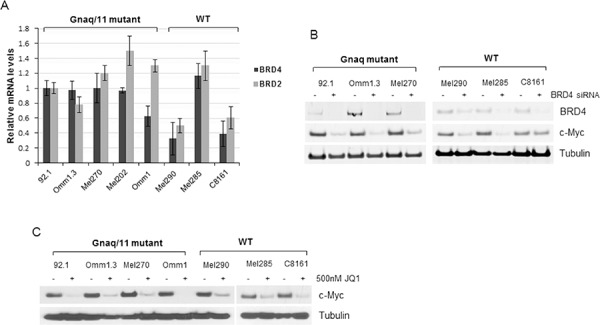
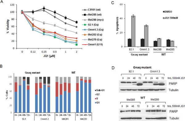
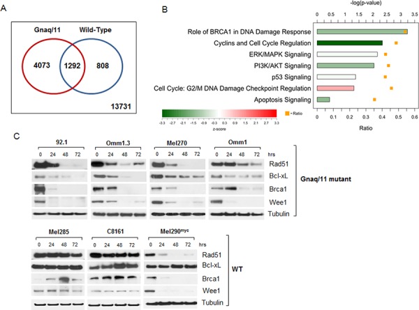
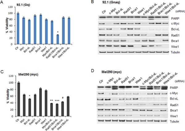
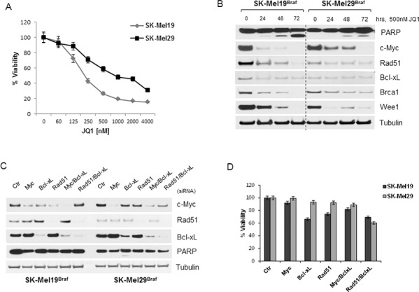
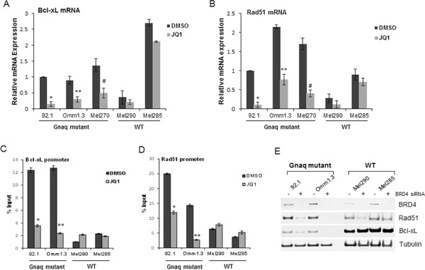
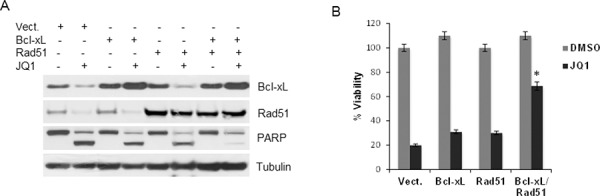
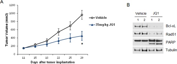
Uveal melanoma (UM) is an aggressive intraocular malignancy with limited therapeutic options. Both primary and metastatic UM are characterized by oncogenic mutations in the G-protein alpha subunit q and 11. Furthermore, nearly 40% of UM has amplification of the chromosomal arm 8q and monosomy of chromosome 3, with consequent anomalies of MYC copy number. Chromatin regulators have become attractive targets for cancer therapy. In particular, the bromodomain and extra-terminal (BET) inhibitor JQ1 has shown selective inhibition of c-Myc expression with antiproliferative activity in hematopoietic and solid tumors. Here we provide evidence that JQ1 had cytotoxic activity in UM cell lines carrying Gnaq/11 mutations, while in cells without the mutations had little effects. Using microarray analysis, we identified a large subset of genes modulated by JQ1 involved in the regulation of cell cycle, apoptosis and DNA repair. Further analysis of selected genes determined that the concomitant silencing of Bcl-xL and Rad51 represented the minimal requirement to mimic the apoptotic effects of JQ1 in the mutant cells, independently of c-Myc. In addition, administration of JQ1 to mouse xenograft models of Gnaq-mutant UM resulted in significant inhibition of tumor growth.Collectively, our results define BRD4 targeting as a novel therapeutic intervention against UM with Gnaq/Gna11 mutations.

Keywords: BRD4; Bcl-xL; Gnaq/11; JQ1; Rad51.


Figures

Figure 1
Figure 1. BRD4 and BRD2 are expressed in UM cells
A. Total RNA was extracted from eight cell lines with the indicated mutational status, and qPCR was performed using gene-specific primers for BRD4 and BRD2. Values were normalized with GAPDH as housekeeping gene using the ΔΔCT method. Values are relative to mRNA levels of 92.1 cells set at 1. Each experiment was performed in triplicates. Bars, mean ± sd. B. BRD4 is active in UM cells. Silencing of BRD4 down-regulates c-Myc expression in Gnaq mutant- and WT cell lines, as detected by immunoblotting. C. Cells were treated with 500 nM JQ1 for 24 hours and cell lysates were analyzed for c-Myc expression and tubulin as loading control.
Figure 2
Figure 2. JQ1 induces cell cycle arrest and apoptosis in UM cells
A. JQ1 reduces viability of a panel of UM cell lines with the indicated mutational status. The cell lines were exposed to 2-fold serial dilutions 2000–100 nM of JQ1 in triplicates for 4 days, and viability was normalized to DMSO-treated cells. Data points are mean ± sd. B. Gnaq-mutant and WT cell lines were treated with DMSO or 500 nM JQ1 over time up to 72 hours. The cells were stained with propidium iodide (PI) and analyzed for cell cycle distribution by flow cytometry. Sub-G1 populations were 19.8% and 19.2% for 92.1 and Omm1.3 cells, respectively. C. UM cells were treated with 500 nM JQ1 for 48 hours, then incubated with YO-PRO dye (green) and PI (red). Bars report the percent of cells with the sum of green and red fluorescence for each condition in triplicates ± sd. D. The same cell lines (Gnaq-mutant top panel; WT, bottom panel) were treated over time with JQ1 and lysed for Western blot analysis, showing induction of apoptosis by PARP cleavage.
Figure 3
Figure 3. Microarray analysis of JQ1-treated cells reveals expression changes of numerous genes involved in signaling pathways, apoptosis and DNA repair
A. Venn-diagram summarizing differentially expressed genes in JQ1-treated Gnaq/11-mutant cell lines (red circle), and WT cells (blue circle), with corresponding overlapping genes as indicated. Data is deposited at GEO accession no. GSE66048. B. Ingenuity Pathway Analysis for genes differentially expressed in Gnaq/11-mutant cells in response to treatment. The bars show the −log(p-value) from a Fisher's Exact Test for enrichment, and the color indicates the z-score for the pathway. The orange squares indicate the ratio of differentially expressed genes in the pathway that were differentially expressed in the mutant cell lines. C. Immunoblot analysis of UM cells with Gnaq/11 mutations (top panel) or without the mutations (WT, lower panel) treated with 500 nM JQ1 over time, using antibodies against the indicated proteins. Each blot is representative of at least 2 experiments showing same results.
Figure 4
Figure 4. Effect of silencing of selected JQ1-regulated genes
The indicated JQ1-regulated genes were silenced in the Gnaq-mutant cell line 92.1 (top) and in the myc-amplified cell line Mel290 (bottom). A. and C. siRNA transfected cells were plated in 96 well plates in triplicates and assayed for cell viability after 72 hours. Viability is calculated as percentage of cells transfected with a control siRNA (Ctr). Graphs are representative of three independent experiments. Bars, mean ± sd. *P < 0.001, **P < 0.01; ***P < 0.05, comparing the effect of gene-specific silencing versus control siRNA-transfected cells. The down-regulation of each gene was analyzed by immunoblotting for 92.1 B. and Mel290 cells D.
Figure 5
Figure 5. BRAF mutant melanoma cells are sensitive to JQ1 through other mechanisms
A. Viability of BRAF-mutant cutaneous melanoma cells (SK-Mel19 and SK-Mel29) was assayed after 4 days of exposure to increasing doses of JQ1 treatments. B. Immunoblot analysis of BRAF-mutant cells treated with 500 nM JQ1 over time, using antibodies against c-Myc, Bcl-xL, Rad51, Brca1, Wee1 and PARP. C. The same genes were silenced and their expression was tested by immunoblotting. D. siRNA-transfected cells were tested for cell viability after 72 hours from transfection. Bars, mean ± sd
Figure 6
Figure 6. JQ1 directly suppresses Bcl-xL and Rad51 in Gnaq/11 mutant cells
The effect of JQ1 on the mRNA of Bcl-xL and Rad51 was confirmed by qPCR in UM cell lines. Total RNA was extracted from cells with different mutational status after 24 h of treatment with 500 nM JQ1, and qPCR was performed using gene-specific primers for Bcl-xL A. and Rad51 B. Values were normalized with GAPDH as housekeeping gene using the ΔΔCT method. Values are relative to mRNA levels of 92.1 untreated cells set at 1. Each experiment was performed two or three times in triplicates. Bars, ± sd. *, **P < 0.01; #P < 0.05, comparing treatment versus DMSO. BRD4 ChIP assay for the Bcl-xL promoter C. and Rad51 promoter D. presented as percent of input, before and after treatment. Bars are representative of two independent experiments ± sd. *P < 0.001; **P < 0.01; #, P < 0.05. E. Downregulation of BRD4 regulates Bcl-xL and Rad51 expression in the Gnaq-mutant cells. The indicated cell lines were transfected with a non-specific siRNA (−) or BRD4 siRNA (+), and analyzed by immunoblotting with the indicated antibodies.
Figure 7
Figure 7. Overexpression of Bcl-xL and Rad51 partially protects cells from JQ1-induced cytotoxic effects
A. 92.1 cells were transfected with an empty vector (pcDNA3), Bcl-xL and Rad51 alone or together, before JQ1 treatment. Cell lysates were subject to immunoblotting using Bcl-xL, Rad51, PARP and tubulin antibodies. B. Viability assay of transfected cells with or without treatment with JQ1. Columns, mean of three independent experiments. ± sd. *P = 0.003 comparing the effect of JQ1 in cells overexpressing Bcl-xL and Rad51 versus vector-transfected cells.
Figure 8
Figure 8. JQ1 inhibits UM tumor growth in vivo
A. JQ1 inhibited tumor growth in a xenograft model with the Gnaq-mutant 92.1 cell line. Six- to eight-week SCID female mice were subcutaneously injected with 92.1 cells. Drug treatments began after tumors reached 100 mm3. Mice bearing tumors were treated daily with JQ1 (35 mg/kg) orally for 5 days each week for a total of 3 weeks. Tumors were measured with calipers every 2 to 3 days and tumor volumes were compared between groups of mice at various points in time. Each value represents the mean measurement of 5 animals, ± SEM, *P < 0.05. B. Xenograft tumors were collected at the end of treatments from two vehicle- and two JQ1-treated mice, and analyzed by Western blotting with antibodies for Bcl-xL, Rad51 and PARP.

References

    1. Dawson MA, Kouzarides T. Cancer Epigenetics: from mechanism to therapy. Cell. 2012;150:12–27. - PubMed
    1. Filippakopoulos P, Qi J, Picaud S, Shen Y, Smith WB, Fedorov O, Morse EM, Keates T, Hickman TT, Felletar I, Philpott M, Munro S, McKeown MR, et al. Selective inhibition of BET bromodomains. Nature. 2010;468:1067–1073. - PMC - PubMed
    1. Fu LL, Tian M, Li X, Li JJ, Huang J, Ouyang L, Zhang Y, Liu B. Inhibition of BET bromodomains as a therapeutic strategy for cancer drug discovery. Oncotarget. 2015;6:5501–5516. - PMC - PubMed
    1. Dawson MA, Prinjha RK, Dittmann A, Giotopoulos G, Bantscheff M, Chan WI, Robson SC, Chung CW, Hopf C, Savitski MM, Huthmacher C, Gudgin E, Lugo D, et al. Inhibition of BET recruitment to chromatin as an effective treatment for MLL-fusion leukaemia. Nature. 2011;478:529–533. - PMC - PubMed
    1. Jang MK, Mochizuki K, Zhou M, Jeong HS, Brady JN, Ozato K. The bromodomain protein Brd4 is a positive regulatory component of P-TEFb and stimulates RNA polymerase II-dependent transcription. Mol Cell. 2005;19:523–534. - PubMed

Publication types

MeSH terms