. 2016 Apr;18(4):488-99.
doi: 10.1111/cmi.12525. Epub 2015 Oct 23.

Vibrio fischeri-derived outer membrane vesicles trigger host development

Affiliations

Vibrio fischeri-derived outer membrane vesicles trigger host development

Marie-Stephanie Aschtgen et al. Cell Microbiol. 2016 Apr.

Abstract

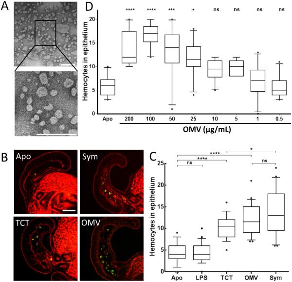
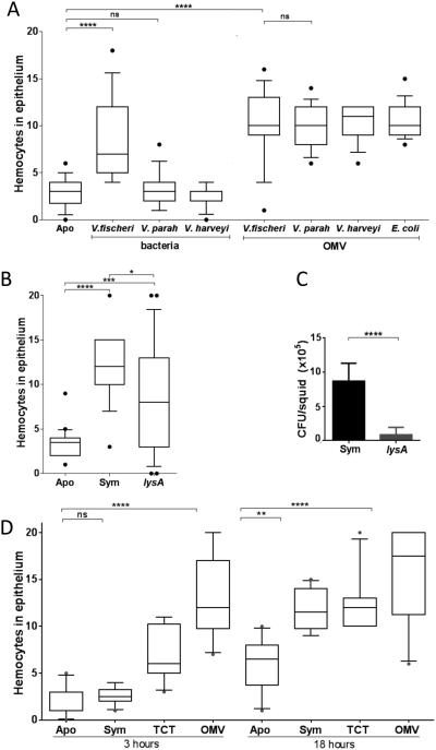
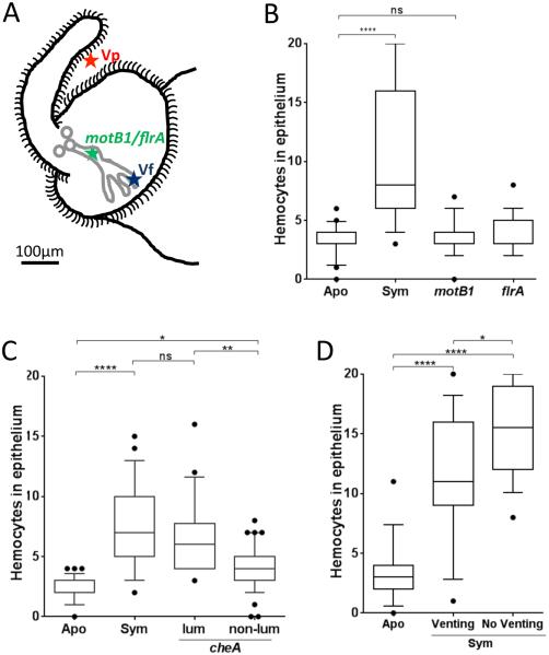
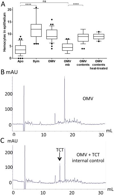
Outer membrane vesicles (OMV) are critical elements in many host-cell/microbe interactions. Previous studies of the symbiotic association between Euprymna scolopes and Vibrio fischeri had shown that within 12 h of colonizing crypts deep within the squid's light organ, the symbionts trigger an irreversible programme of tissue development in the host. Here, we report that OMV produced by V. fischeri are powerful contributors to this process. The first detectable host response to the OMV is an increased trafficking of macrophage-like cells called haemocytes into surface epithelial tissues. We showed that exposing the squid to other Vibrio species fails to induce this trafficking; however, addition of a high concentration of their OMV, which can diffuse into the crypts, does. We also provide evidence that tracheal cytotoxin released by the symbionts, which can induce haemocyte trafficking, is not part of the OMV cargo, suggesting two distinct mechanisms to induce the same morphogenesis event. By manipulating the timing and localization of OMV signal delivery, we showed that haemocyte trafficking is fully induced only when V. fischeri, the sole species able to reach and grow in the crypts, succeeds in establishing a sustained colonization. Further, our data suggest that the host's detection of OMV serves as a symbiotic checkpoint prior to inducing irreversible morphogenesis.

Keywords: OMV; euprymna scolopes; haemocyte; immune response; symbiosis.


Figures

Fig. 1
Fig. 1
OMV induce a phenotype associated with PG-linked light-organ morphogenesis. Influence of OMV addition on hemocyte trafficking. (A) Negative-stained TEM of purified OMV produced by wild-type V. fischeri ES114; boxed area enlarged below. Scale bars indicate 200 nm. (B) Confocal micrographs of one appendage of a juvenile light organ: red, rhodamine phalloidin (filamentous actin); green, DNAse I (hemocytes). Scale bar = 50 μm. (C) Quantification of hemocyte trafficking in symbiotic animals (exposed to 104 V. fischeri cfu/mL), or in animals treated with either TCT (1 μM), LPS (10 μg/mL) or OMV (100 μg of protein/mL), after 18 h; these levels of TCT and LPS are in the range that elicits other host responses (Foster et al. 2000; Koropatnick et al. 2004). Hemocytes were counted in the sinuses of the anterior appendage of one epithelial field per light organ. n=20; One-way ANOVA analysis (F=37; p<0.0001). (D) Levels of hemocyte trafficking in the anterior appendages of animals exposed for 18 h to a range of OMV concentrations, from 0.5 to 200 μg of protein/mL. n=20. One-way ANOVA analysis (F=28; p<0).
Fig. 2
Fig. 2
Hemocyte trafficking is dependent on the intensity and location of signal delivery. The extent of trafficking was determined by counting hemocytes in the anterior appendage of one epithelial field per light organ. (A) Juvenile squid were exposed for 18 h to either 104 cfu of V. fischeri, V. parahaemolyticus (V. parah), or V. harveyi per ml, or 100 μg of OMV produced by these strains, or by E. coli. One-way ANOVA analysis (F=21; p<0.0001). (B) Juvenile squid were exposed to 104 cfu of wild-type V. fischeri (Sym), or a isogenic lysA derivative, per ml. One-way ANOVA analysis (F=20; p<0.0001). (C) Symbiont population levels after 48 h in squid exposed to either the wild-type (Sym) or the auxotrophic lysA strain, determined in three independent experiments, representing a total of 60 squids per condition. Starting inoculum levels ranged from 8-11 × 103 cfu/mL. Graphical and errors bars indicate average and standard deviation of data. p<0.05. (D) Juvenile squid were exposed for either 3 or 18 h to seawater only (Apo), or seawater containing 104 V. fischeri cfu/mL (Sym), or to 1 μM TCT, or 100 μg of OMV protein/mL. One-way ANOVA analysis (F=27; p<0.0001).
Fig. 3
Fig. 3
Bacterial delivery of the hemocyte-trafficking signal. The extent of hemocyte trafficking was determined by counting hemocytes in the sinuses of the anterior appendage of one epithelial field per light organ. (A) Diagram of the left side of a juvenile light organ showing both the surface in contact with the seawater (black outline) and the internal structures (pores, ducts, antechamber and deep crypts) through which symbionts migrate (gray). Indicated are the limits of migration of V. parahaemolyticus (Vp), which like other non-symbionts doesn’t enter the three pores; V. fischeri wild type (Vf), which migrates to and grows in the deep crypts; and, motility mutants (flrA; motB1), which cannot pass beyond the organ’s antechamber. The localization of the cheA mutant, which has been found to stochastically enter the pore (Mandel et al. 2012), is complex and has not been indicated here. When added to the surrounding seawater small particles, like OMVs, can diffuse into the crypts. See text for a full explanation. (B) Juvenile squid were exposed to motB1 and flrA mutants unable to complete the migration into the crypts. (C) Juvenile squid were exposed to 104 cfu of a V. fischeri cheA mutant per mL. After 18 h, animals were sorted into two groups: detectably luminescent (Lum) or not (Non-lum); in the former group, the cheA cells were presumed to have reached a crypt and proliferated, while in the latter, the cheA cells were presumed to have been unable to establish a sustained colonization. n=60. One-way ANOVA analysis (F=31; p<0.0001). (D) After inoculation, animals were either kept 12 h in the dark, followed by 4 h in the light to induce venting through the ducts and pores (Venting), or kept 16 h in the dark (No Venting) before the level of hemocyte trafficking was determined. One-way ANOVA analysis (F=37; p<0.0001).
Fig. 4
Fig. 4
OMV induce hemocyte infiltration, but do not contain TCT. (A) To quantify hemocyte trafficking, 30 animals were exposed either to 104 V. fischeri cfu/mL (Sym), or to purified OMV (100 μg/mL), OMV membranes (mb), OMV contents, or heat-treated OMV contents. Lipid and protein concentrations after fractionation were determined by the FM4-based fluorescence assay and Qubit. Fluorescence associated with the OMV content is 8% and 92% with OMV membranes. One-way ANOVA analysis (F=34; p<0.0001). (B-C) Evidence for the presence of TCT in OMV contents was determined by reversed-phase HPLC. (B) HPLC profile of OMV sample (100 μg of protein). (C) Internal control of 10 nmole of TCT was loaded with the same OMV sample.
Fig. 5
Fig. 5
Internalization of V. fischeri OMV. (A) Confocal-microscopy sections showing the appearance of OMV in the cytoplasm of epithelial cells of the light-organ appendages after 3 h incubation with: FITC-labeled (green) OMV (OMV), OMV and cytochalasin D (OMV + cytD), or 2-μm diameter FITC-labeled beads (beads). Nuclei were stained with TOTO3 (blue), and F-actin with rhodamine phalloidin (red). (B) FITC-labeled OMV were incubated with hemocytes, and their fluorescence was measured over time as an estimation of OMV internalization. Data are presented as mean fluorescence intensity (FI), and error bars indicate one standard deviation; p < 0.05. (C) The numbers of cells with internalized FITC-labeled OMV or beads were determined for 30 treated hemocytes by confocal imaging. Data analyzed with one-way ANOVA analysis of differences: (**), p < 0.001. (D) Confocal-microscopy sections illustrating the internalization of FITC-labeled OMV by isolated hemocytes 90 min after an in vitro incubation; staining as in (A). A z-stack of sections illustrates internalized OMV fluorescence in the OMV-treated, but not control hemocytes. (E) Effect of cytochalasin D pretreatment on induction of hemocyte trafficking by OMV or TCT. n=20; One-way ANOVA analysis (F=18; p<0.0001). Size bars = 5 μm.

References

    1. Adin DM, Engle JT, Goldman WE, McFall-Ngai MJ, Stabb EV. Mutations in ampG and lytic transglycosylase genes affect the net release of peptidoglycan monomers from Vibrio fischeri. J Bacteriol. 2009;191:2012–2022. - PMC - PubMed
    1. Bertani G. Studies on lysogenesis. I. The mode of phage liberation by lysogenic Escherichia coli. J Bacteriol. 1951;62:293–300. - PMC - PubMed
    1. Bielig H, Rompikuntal PK, Dongre M, Zurek B, Lindmark B, Ramstedt M, et al. NOD-like receptor activation by outer membrane vesicles from Vibrio cholerae Non-O1 Non-O139 strains is modulated by the quorum-sensing regulator HapR. Infect Immun. 2011;79:1418–1427. - PMC - PubMed
    1. Boettcher KJ, Ruby EG. Depressed light emission by symbiotic Vibrio fischeri of the sepiolid squid Euprymna scolopes. J Bacteriol. 1990;172:3701–3706. - PMC - PubMed
    1. Boudreau MA, Fisher JF, Mobashery S. Messenger functions of the bacterial cell wall-derived muropeptides. Biochemistry. 2012;51:2974–2990. - PMC - PubMed

Publication types

LinkOut - more resources