. 2015 Nov 20;290(47):28097-28106.
doi: 10.1074/jbc.M115.680090. Epub 2015 Sep 23.

Ablation of the Ferroptosis Inhibitor Glutathione Peroxidase 4 in Neurons Results in Rapid Motor Neuron Degeneration and Paralysis

Affiliations

Ablation of the Ferroptosis Inhibitor Glutathione Peroxidase 4 in Neurons Results in Rapid Motor Neuron Degeneration and Paralysis

Liuji Chen et al. J Biol Chem. .

Abstract

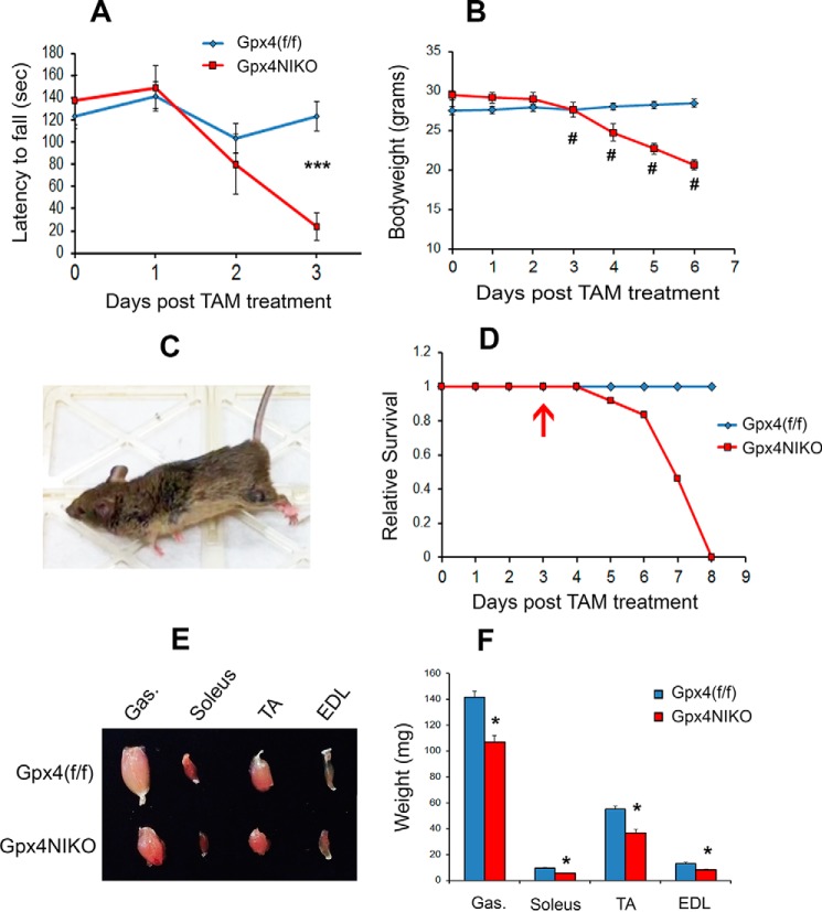
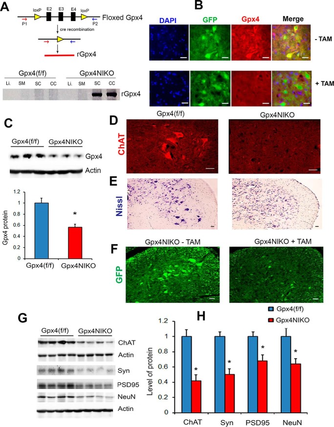
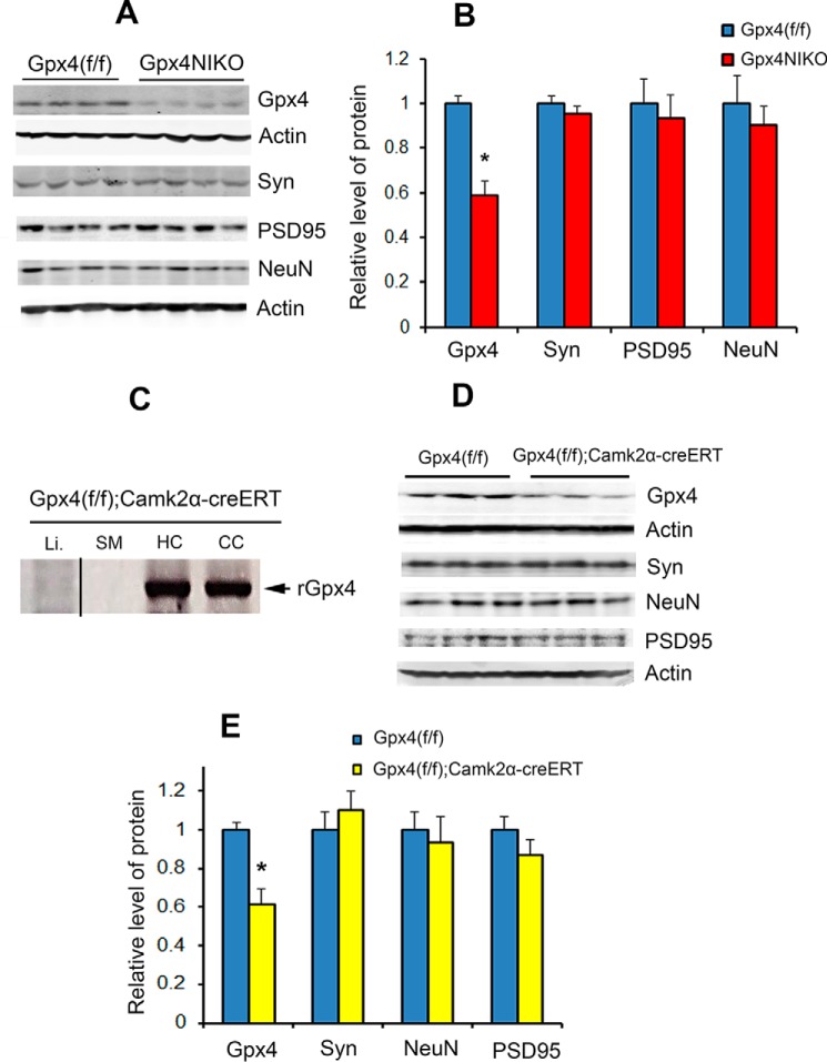
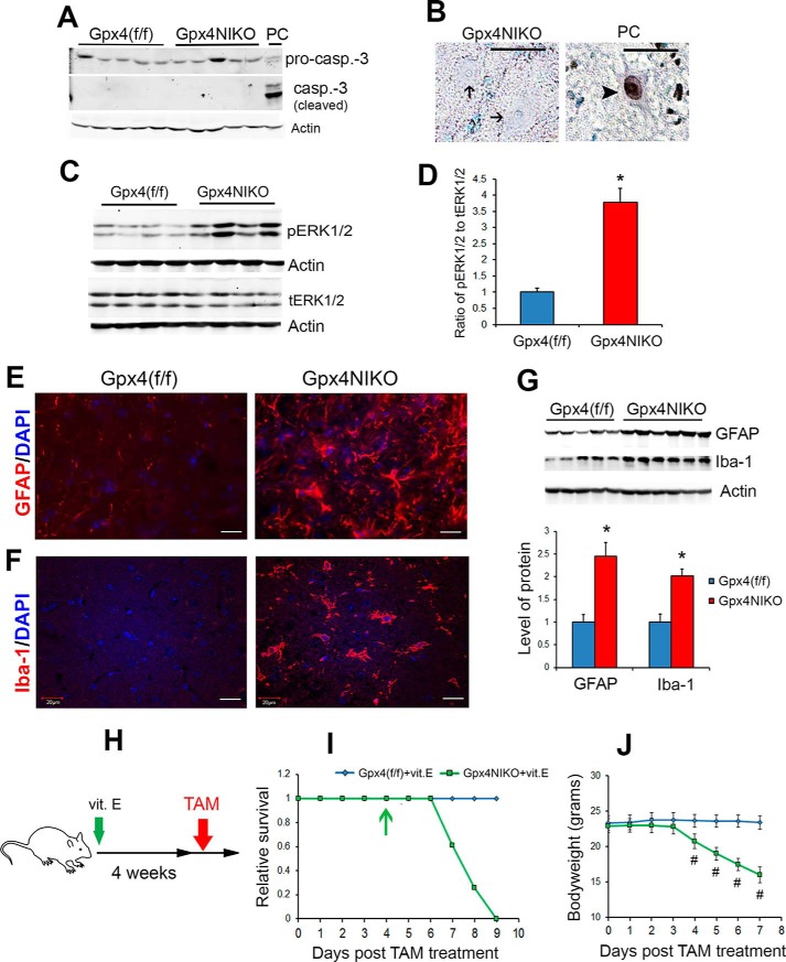
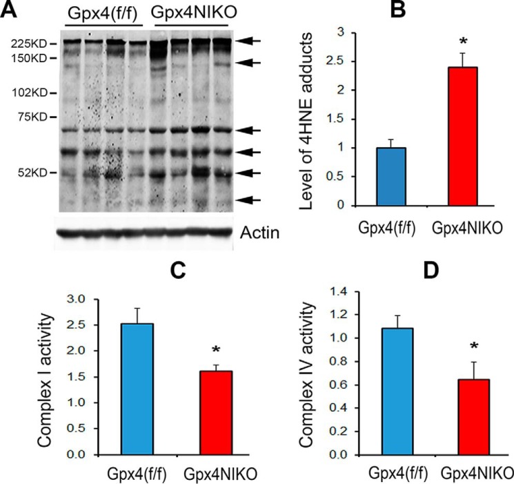
Glutathione peroxidase 4 (GPX4), an antioxidant defense enzyme active in repairing oxidative damage to lipids, is a key inhibitor of ferroptosis, a non-apoptotic form of cell death involving lipid reactive oxygen species. Here we show that GPX4 is essential for motor neuron health and survival in vivo. Conditional ablation of Gpx4 in neurons of adult mice resulted in rapid onset and progression of paralysis and death. Pathological inspection revealed that the paralyzed mice had a dramatic degeneration of motor neurons in the spinal cord but had no overt neuron degeneration in the cerebral cortex. Consistent with the role of GPX4 as a ferroptosis inhibitor, spinal motor neuron degeneration induced by Gpx4 ablation exhibited features of ferroptosis, including no caspase-3 activation, no TUNEL staining, activation of ERKs, and elevated spinal inflammation. Supplementation with vitamin E, another inhibitor of ferroptosis, delayed the onset of paralysis and death induced by Gpx4 ablation. Also, lipid peroxidation and mitochondrial dysfunction appeared to be involved in ferroptosis of motor neurons induced by Gpx4 ablation. Taken together, the dramatic motor neuron degeneration and paralysis induced by Gpx4 ablation suggest that ferroptosis inhibition by GPX4 is essential for motor neuron health and survival in vivo.

Keywords: GPX4; cell death; ferroptosis; gene knockout; lipid peroxidation; motor neurons; muscle atrophy; neurodegeneration; paralysis.


Figures

FIGURE 1.
FIGURE 1.
Gpx4 ablation induced rapid paralysis, muscle atrophy, and death in Gpx4NIKO mice, a mouse model with inducible ablation of Gpx4 in neurons. A, rotarod performance of Gpx4NIKO mice (n = 7, male and female) and Gpx4(f/f) mice (n = 7, male and female) after TAM treatment. ***, p < 0.001. B, body weights of Gpx4NIKO mice (n = 8, male) and Gpx4(f/f) mice (n = 11, male) after TAM treatment. #, p < 0.01. C, a paralyzed Gpx4NIKO mouse on day 6 post-TAM treatment. D, survival curves of Gpx4NIKO mice (n = 24, male and female) and Gpx4(f/f) mice (n = 16, male and female) after TAM treatment. The arrow indicates onset of paralysis. E, individual hind limb muscles (gastrocnemius (Gas), extensor digitorum longus (EDL), soleus, and tibialis anterior (TA)) from a Gpx4NIKO mouse and a Gpx4(f/f) mouse on day 6 post-TAM treatment. F, hind limb muscle weights on day 6 post-treatment (n = 5, male). *: p < 0.05.
FIGURE 2.
FIGURE 2.
A dramatic degeneration of spinal cord motor neurons in the paralyzed Gpx4NIKO mice. A, detection of the recombined Gpx4 gene allele (rGpx4) by PCR only in nervous tissues from TAM-treated Gpx4NIKO mice. SC, spinal cord; CC, cerebral cortex; SM, skeletal muscle; Li, liver. B, spinal cord sections from control Gpx4NIKO mice without TAM treatment (− TAM) and TAM-treated Gpx4NIKO mice (+ TAM), showing reduced GPX4 immunofluorescence in spinal neurons of Gpx4NIKO mice. C, representative Western blots of GPX4 protein in spinal cord tissues and quantified GPX4 protein levels in spinal cord tissues (n = 9, male and female) for both groups. *, p < 0.05. D, lumbar spinal cord sections from Gpx4(f/f) mice and Gpx4NIKO mice on day 6 post-TAM treatment, showing ChAT-positive motor neurons. E, Nissl-stained lumbar spinal cord sections from Gpx4(f/f) mice and Gpx4NIKO mice. F, lumbar spinal cord sections from control Gpx4NIKO mice (− TAM) and Gpx4NIKO mice with TAM treatment (+ TAM), showing GFP-positive spinal cord neurons. G, representative Western blots showing levels of ChAT, synaptophysin (Syn), PSD95, and NeuN in spinal cord tissues from Gpx4NIKO mice and Gpx4(f/f) mice. H, quantified results of ChAT, Syn, PSD95, and NeuN levels in spinal cords from Gpx4NIKO mice and Gpx4(f/f) mice (n = 9, male and female). *, p < 0.05. Scale bars = 20 μm.
FIGURE 3.
FIGURE 3.
Gpx4 ablation resulted in no overt neurodegeneration in the cerebral cortex. A, Western blots showing levels of GPX4, Syn, NeuN, and PSD95 in cortex tissues from Gpx4(f/f) mice and Gpx4NIKO mice on day 6 post TAM treatment (n = 9, male and female). B, quantified levels of GPX4, Syn, NeuN, and PSD95 in cortex tissues. *, p < 0.05. C, detection of rGpx4 by PCR only in nervous tissues from TAM-treated Gpx4(f/f);Camk2α-creERT mice. The image was assembled from different parts of a gel. CC, cerebral cortex; HC, hippocampus; SM, skeletal muscle; Li, liver. D, Western blots showing levels of GPX4, Syn, NeuN, and PSD95 in cortex tissues from Gpx4(f/f) mice and Gpx4(f/f);Camk2α-creERT mice 2 weeks post-TAM treatment. E, quantified levels of GPX4, Syn, NeuN, and PSD95 in cortex tissues from Gpx4(f/f) mice and Gpx4(f/f);Camk2α-creERT mice 2 weeks post-TAM treatment (n = 3, male and female). *, p < 0.05.
FIGURE 4.
FIGURE 4.
Motor neuron degeneration in Gpx4NIKO mice exhibited features of ferroptosis. A, Western blots showing spinal proteins probed with an antibody against pro-caspase 3 (pro-casp.-3) and activated caspase-3 (casp.-3 (cleaved)). Tissues were obtained at day 3 post TAM treatment. PC, positive control. B, spinal motor neurons from Gpx4NIKO mice on day 3 post-TAM treatment stained by TUNEL. Arrows indicate TUNEL-negative nuclei. PC, positive control (a motor neuron treated with nuclease); arrowhead, a TUNEL-positive nucleus. C, Western blots showing levels of phosphorylated ERK1/2 (pERK1/2) and total ERK1/2 (tERK1/2) on day 6 post-TAM treatment. D, quantified ratios of pERK1/2 to tERK1/2. E, lumbar spinal cord sections from Gpx4(f/f) mice and Gpx4NIKO mice on day 6 post-TAM treatment stained with an anti-GFAP antibody. *, p < 0.05. F, lumbar spinal cord sections from Gpx4(f/f) mice and Gpx4NIKO mice stained with an anti-Iba-1 antibody. G, Western blots showing levels of GFAP and Iba-1 in spinal cord tissues from Gpx4(f/f) mice and Gpx4NIKO mice (day 6) and quantified results of GFAP and Iba-1 levels (n = 5–6, male and female). *, p < 0.05. H, design of the vitamin E (vit. E) supplement study. I, survival curves of vitamin E-fed Gpx4NIKO mice (Gpx4NIKO+vit.E, n = 23, male and female) and Gpx4(f/f) mice (Gpx4(f/f)+vit.E, n = 22, male and female) after TAM treatment. The arrow indicates onset of paralysis. J, body weights of vitamin E-fed Gpx4NIKO mice and Gpx4(f/f) mice after TAM treatment. #, p < 0.01. Scale bars = 20 μm.
FIGURE 5.
FIGURE 5.
Increased lipid peroxidation and mitochondrial dysfunction at onset of paralysis in TAM-treated Gpx4NIKO mice. A, Western blots of spinal cord proteins probed with an anti-4-HNE antibody and then with an anti-actin antibody to control loading. Arrows indicate 4-HNE protein adducts. Tissues were obtained on day 3 post TAM treatment. B, quantified results of 4-HNE adducts (n = 4, male and female). *, p < 0.05. C, activity of electron transport chain complex I of spinal cord mitochondria on day 3. *, p < 0.05. D, activity of electron transport chain complex IV of spinal cord mitochondria on day 3 (n = 5). *, p < 0.05.

References

    1. Brigelius-Flohé R. (1999) Tissue-specific functions of individual glutathione peroxidases. Free Radic. Biol. Med. 27, 951–965 - PubMed
    1. Imai H., and Nakagawa Y. (2003) Biological significance of phospholipid hydroperoxide glutathione peroxidase (PHGPx, GPx4) in mammalian cells. Free Radic. Biol. Med. 34, 145–169 - PubMed
    1. Ran Q., Van Remmen H., Gu M., Qi W., Roberts L. J. 2nd, Prolla T., and Richardson A. (2003) Embryonic fibroblasts from Gpx4+/− mice: a novel model for studying the role of membrane peroxidation in biological processes. Free Radic. Biol. Med. 35, 1101–1109 - PubMed
    1. Ran Q., Liang H., Gu M., Qi W., Walter C. A., Roberts L. J. 2nd, Herman B., Richardson A., and Van Remmen H. (2004) Transgenic mice overexpressing glutathione peroxidase 4 are protected against oxidative stress-induced apoptosis. J. Biol. Chem. 279, 55137–55146 - PubMed
    1. Yang W. S., and Stockwell B. R. (2008) Synthetic lethal screening identifies compounds activating iron-dependent, nonapoptotic cell death in oncogenic-RAS-harboring cancer cells. Chem. Biol. 15, 234–245 - PMC - PubMed

Publication types

Substances