. 2016 Feb;183(2):280-93.
doi: 10.1111/cei.12710. Epub 2015 Nov 17.

Modulating of ocular inflammation with macrophage migration inhibitory factor is associated with notch signalling in experimental autoimmune uveitis

Affiliations

Modulating of ocular inflammation with macrophage migration inhibitory factor is associated with notch signalling in experimental autoimmune uveitis

H Yang et al. Clin Exp Immunol. 2016 Feb.

Abstract

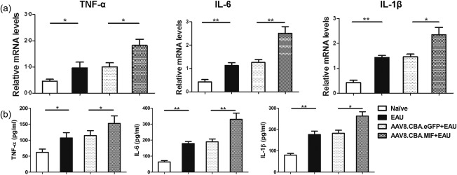
The aim of this study was to examine whether macrophage migration inhibitory factor (MIF) could exaggerate inflammatory response in a mouse model of experimental autoimmune uveitis (EAU) and to explore the underlying mechanism. Mutant serotype 8 adeno-associated virus (AAV8) (Y733F)-chicken β-actin (CBA)-MIF or AAV8 (Y733F)-CBA-enhanced green fluorescent protein (eGFP) vector was delivered subretinally into B10.RIII mice, respectively. Three weeks after vector delivery, EAU was induced with a subcutaneous injection of a mixture of interphotoreceptor retinoid binding protein (IRBP) peptide with CFA. The levels of proinflammatory cytokines were detected by real-time polymerase chain reaction (PCR) and enzyme-linked immunosorbent assay (ELISA). Retinal function was evaluated with electroretinography (ERG). We found that the expression of MIF and its two receptors CD74 and CD44 was increased in the EAU mouse retina. Compared to AAV8.CBA.eGFP-injected and untreated EAU mice, the level of proinflammatory cytokines, the expression of Notch1, Notch4, delta-like ligand 4 (Dll4), Notch receptor intracellular domain (NICD) and hairy enhancer of split-1 (Hes-1) increased, but the ERG a- and b-wave amplitudes decreased in AAV8.CBA.MIF-injected EAU mice. The Notch inhibitor N-[N-(3,5-difluorophenacetyl)-l-alanyl]-S-phenylglycine t-butyl ester (DAPT) reduced the expression of NICD, Hes-1 and proinflammatory cytokines. Further, a MIF antagonist ISO-1 attenuated intraocular inflammation, and inhibited the differentiation of T helper type 1 (Th1) and Th17 in EAU mice. We demonstrated that over-expression of MIF exaggerated ocular inflammation, which was associated with the activation of the Notch signalling. The expression of both MIF and its receptors are elevated in EAU mice. Over-expression of MIF exaggerates ocular inflammation, and this exaggerated inflammation is associated with the activation of the Notch signalling and Notch pathway. Our data suggest that the MIF-Notch axis may play an important role in the pathogenesis of EAU. Both the MIF signalling pathways may be promising targets for developing novel therapeutic interventions for uveitis.

Keywords: EAU; MIF; Notch; inflammation; retina.


Figures

Figure 1
Figure 1
Expression of macrophage migration inhibitory factor (MIF) and its two receptors in the retina of experimental autoimmune uveitis (EAU) mice at day 14 post‐immunization. EAU was induced in B10.RIII mice by active immunization with a mixture of interphotoreceptor retinoid binding protein (IRBP) peptide with complete Freund's adjuvant (CFA). The eyeballs were collected 14 days later to evaluate the expression of MIF, CD74 and CD44. (a) Representative images of immunostaining showed MIF‐positive cells (brown) in naive mice (left panel), EAU mice (middle panel) and negative control (right panel). Real‐time polymerase chain reaction (PCR) showed that the levels of MIF (b), CD74 (c) and CD44 (d) mRNAs increased in EAU than in naive mice. The relative gene expression was normalized to glyceraldehyde‐3‐phosphate dehydrogenase (GAPDH). Data are mean ± standard error of the mean (s.e.m.) and were representative of four independent experiments. **P < 0·01; ***P < 0·001. The scale bar represents 50 µm. Six mice were used in each group.
Figure 2
Figure 2
Validation of serotype 8 adeno‐associated virus [AAV8(Y733F)]‐mediated enhanced green fluorescent protein (eGFP) reporter gene expression in mouse retina. (a) One week after subretinal injection of AAV8.chicken β‐actin (CBA).eGFP, the white arrow indicates the injection area. (b) Three weeks after subretinal injection of AAV8.CBA.eGFP, GFP expression was detected by fundus examination. Delivery of the vector (1 μl) led to a wide range of transduction. (d) Higher transduction of GFP was seen in the photoreceptor and in the RPE layers. (c) The control image of GFP expression was examined under white light by fluorescence microscopy. Green = GFP; RPE = retinal pigment epithelium; ONL = outer nuclear layer.
Figure 3
Figure 3
Retinal protein expression of macrophage migration inhibitory factor (MIF) in serotype 8 adeno‐associated virus (AAV8).chicken β‐actin (CBA).MIF‐injected experimental autoimmune uveitis (EAU) mice. MIF protein level was higher in AAV8.CBA.MIF than in the AAV8.CBA.enhanced green fluorescent protein (eGFP)‐injected eyes. The relative expression was normalized to β‐actin. Data are mean ± standard error of the mean (s.e.m.) and were representative of three independent experiments. *P < 0·05. Six mice were used in each group.
Figure 4
Figure 4
Expression of proinflammatory cytokines in macrophage migration inhibitory factor (MIF) transduced experimental autoimmune uveitis (EAU) mice. Retinal mRNA and protein were measured at day 14 after immunization with a mixture of interphotoreceptor retinoid binding protein (IRBP) peptide with complete Freund's adjuvant (CFA). (a) Real‐time polymerase chain reaction (PCR) showed increase of proinflammatory cytokines [tumour necrosis factor (TNF)‐α, interleukin (IL)−1β and IL‐6] in AAV8.CBA.MIF‐injected EAU mice. (b) TNF‐α, IL‐1β and IL‐6 levels increased in AAV8.chicken β‐actin (CBA).MIF‐injected EAU mice than in AAV8.CBA.enhanced green fluorescent protein (eGFP)‐injected EAU mice. The relative gene expression was normalized to glyceraldehyde‐3‐phosphate dehydrogenase (GAPDH). Data are mean ± standard error of the mean (s.e.m.) and were representative of three to four independent experiments. *P < 0·05; ***P < 0·001. Six mice were used in each group.
Figure 5
Figure 5
The effect of macrophage migration inhibitory factor (MIF) on retinal function in experimental autoimmune uveitis (EAU) mice. Retinal functions were evaluated by electroretinography (ERG) at day 14 after immunization with a mixture of interphotoreceptor retinoid binding protein (IRBP) peptide with complete Freund's adjuvant (CFA). (a) Representative ERG responses in the naive mice, AAV8.chicken β‐actin (CBA).enhanced green fluorescent protein (eGFP)‐injected EAU mice and AAV8.CBA.MIF‐injected EAU mice. (b) ERG amplitudes versus flash intensity for dark‐adapted a‐wave, b‐wave and light‐adapted b‐wave. Dark‐adapted amplitudes of ERG a‐wave and b‐wave, light‐adapted amplitudes of ERG b‐wave decreased in the EAU mice. MIF exaggerated the functional damage in EAU mice. Data are presented as mean ± standard error of the mean (s.e.m.) of 12 mice. *P < 0·05 versus naive group; **P < 0·01, ***P < 0·001, # P < 0·05 versus EAU group; ## P < 0·01; ### P < 0·001.
Figure 6
Figure 6
Expression of Notch signal pathway in macrophage migration inhibitory factor (MIF) over‐expressed experimental autoimmune uveitis (EAU) mice. (a) Expression of Notch receptors and their ligands in the retina of EAU. The levels of Notch1 and delta‐like ligand 4 (Dll4) increased in EAU mice at day 14 after immunization with a mixture of interphotoreceptor retinoid binding protein (IRBP) peptide with complete Freund's adjuvant (CFA). (b) Expression of Notch receptors and their ligands in the AAV8.chicken β‐actin (CBA).MIF‐injected EAU mice. The expression of Notch1, Notch4 and Dll4 was higher in the AAV8.CBA.MIF‐injected EAU mice than in the AAV8.CBA.enhanced green fluorescent protein (eGFP)‐injected EAU mice. (c) Protein expression of Notch receptor intracellular domain (NICD) in the AAV8.CBA.MIF‐injected EAU mice. NICD protein level was higher in the AAV8.CBA.MIF‐injected eyes than in the AAV8.CBA.eGFP‐injected eyes. (d) Protein expression of hairy enhancer of split‐1 (Hes‐1) in the AAV8.CBA.MIF‐injected EAU mice. Hes‐1 protein level increased in the AAV8.CBA.MIF‐treated eyes compared with the AAV8.CBA.eGFP‐injected eyes. The relative expression was normalized to β‐actin. Data are mean ± standard error of the mean (s.e.m.) and were representative of three independent experiments. *P < 0·05. Six mice were used in each group.
Figure 7
Figure 7
Expression of proinflammatory cytokines in the N‐[N‐(3,5‐difluorophenacetyl)‐l‐alanyl]‐S‐phenylglycine t‐butyl ester (DAPT)‐treated AAV8.CBA.macrophage migration inhibitory factor (MIF)‐injected experimental autoimmune uveitis (EAU) mice. Retinal mRNA and protein were examined at day 14 after a mixture of interphotoreceptor retinoid binding protein (IRBP) peptide with complete Freund's adjuvant (CFA) immunization. Real‐time polymerase chain reaction (PCR) showed increased level of proinflammatory cytokines [tumour necrosis factor (TNF)‐α, interleukin (IL)‐1β, IL‐17 and interferon (IFN)‐γ] in the AAV8.CBA.MIF‐injected EAU eyes. The relative gene expression was normalized to glyceraldehyde‐3‐phosphate dehydrogenase (GAPDH). Data are mean ± standard error of the mean (s.e.m.) and were representative of three to four independent experiments. *P < 0·05. Six mice were used in each group.
Figure 8
Figure 8
The severity of histological score and the expression of proinflammatory cytokines in the ISO‐1‐treated experimental autoimmune uveitis (EAU) mice. Histological scores were assessed with haematoxylin and eosin (H&E) staining at day 14 after immunization. Retinal damage was ameliorated in the ISO‐1‐treated EAU mice (b). Representative retinal histological images of the vehicle‐treated and the ISO‐1‐treated EAU mice (a). Inflammatory cellular infiltration and retinal folds were evident in the vehicle‐treated EAU mice, but was not seen in the ISO‐1‐treated groups. The levels of proinflammatory cytokines [tumour necrosis factor (TNF)‐α, interleukin (IL)‐1β, IL‐17 and interferon (IFN)‐γ] in the ISO‐1‐treated EAU mice were reduced (c). The scale bar represents 50 µm. Data are shown as mean ± standard error of the mean (s.e.m.). *P < 0·05.
Figure 9
Figure 9
The differentiation of T helper type 1 (Th1) and Th17 in the ISO‐1‐treated experimental autoimmune uveitis (EAU) mice. ISO‐1 reduced the proportions of Th1 and Th17 cells expressing interferon (IFN)‐γ, interleukin (IL)‐17 in EAU at day 14. Lymphocytes from the vehicle and the ISO‐1‐treated EAU mice were collected at day 14 post‐immunization and the frequency of IFN‐γ+CD4+ and IL‐17+CD4+ T cells were tested by flow cytometry. (a,c) Representative intracellular expression of IFN‐γ and IL‐17, respectively. (b,d) The averages of four independent experiments presented as the percentages of IFN‐γ‐ and IL‐17‐positive cells in the lymphocytes. Numbers represent percentage of positive cells. Data are mean ± standard error of the mean (s.e.m.) . *P < 0·05. Six mice were used in each group.

References

    1. George RK, Chan CC, Whitcup SM, Nussenblatt RB. Ocular immunopathology of Behcet's disease. Surv Ophthalmol 1997; 42:157–62. - PubMed
    1. Singh VK, Biswas S, Anand R, Agarwal SS. Experimental autoimmune uveitis as animal model for human posterior uveitis. Indian J Med Res 1998; 107:53–67. - PubMed
    1. Caspi RR. A look at autoimmunity and inflammation in the eye. J Clin Invest 2010; 120:3073–83. - PMC - PubMed
    1. Calandra T, Roger T. Macrophage migration inhibitory factor: a regulator of innate immunity. Nat Rev Immunol 2003; 3:791–800. - PMC - PubMed
    1. Roger T, David J, Glauser MP, Calandra T. MIF regulates innate immune responses through modulation of Toll‐like receptor 4. Nature 2001; 414:920–4. - PubMed

Publication types

MeSH terms