. 2015 Sep;5(9):150118.
doi: 10.1098/rsob.150118.

The human parasite Leishmania amazonensis downregulates iNOS expression via NF-κB p50/p50 homodimer: role of the PI3K/Akt pathway

Affiliations

The human parasite Leishmania amazonensis downregulates iNOS expression via NF-κB p50/p50 homodimer: role of the PI3K/Akt pathway

Teresa C Calegari-Silva et al. Open Biol. 2015 Sep.

Abstract

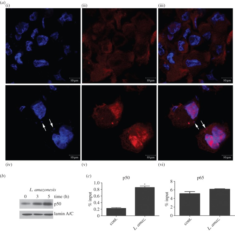
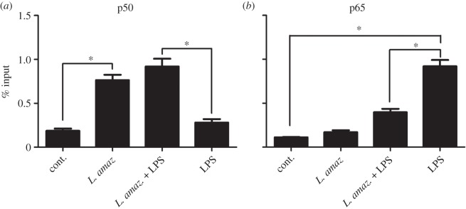
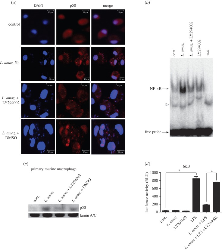
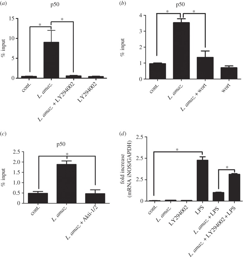
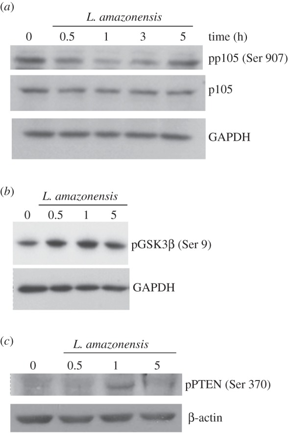
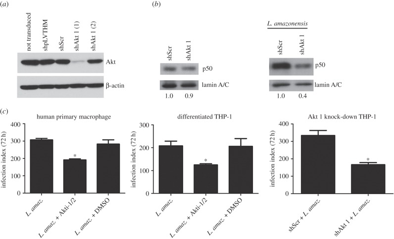
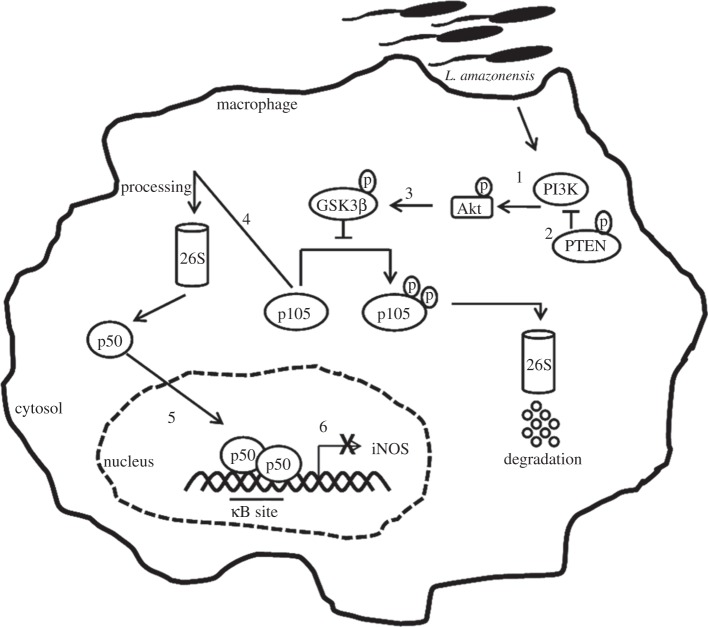
Leishmania amazonensis activates the NF-κB transcriptional repressor homodimer (p50/p50) and promotes nitric oxide synthase (iNOS) downregulation. We investigated the role of PI3K/Akt in p50/p50 NF-κB activation and the effect on iNOS expression in L. amazonensis infection. The increased occupancy of p50/p50 on the iNOS promoter of infected macrophages was observed and we demonstrated that both p50/p50 NF-κB induction and iNOS downregulation in infected macrophages depended on PI3K/Akt activation. Importantly, the intracellular growth of the parasite was also impaired during PI3K/Akt signalling inhibition and in macrophages knocked-down for Akt 1 expression. It was also observed that the increased nuclear levels of p50/p50 in L. amazonensis-infected macrophages were associated with reduced phosphorylation of 907 Ser p105, the precursor of p50. Corroborating these data, we demonstrated the increased levels of phospho-9 Ser GSK3β in infected macrophages, which is associated with GSK3β inhibition and, consequently, its inability to phosphorylate p105. Remarkably, we found that the levels of pPTEN 370 Ser, a negative regulator of PI3K, increased due to L. amazonensis infection. Our data support the notion that PI3K/Akt activity is sustained during the parasite infection, leading to NF-κB 105 phosphorylation and further processing to originate p50/p50 homodimers and the consequent downregulation of iNOS expression.

Keywords: Leishmania; PI3K/Akt; macrophage; nuclear factor kappa B.


Figures

Figure 1.
Figure 1.
Leishmania amazonensis increases NF-κB/p50 subunit nuclear levels and the occupancy on the iNOS promoter. THP-1 cells were differentiated into macrophages and infected with promastigotes of L. amazonensis at the indicated times. (a) Confocal photomicrographs from control (i)(iii)) and L. amazonensis 5 h infection (iv)(vi)) immunostained with p50 (red) and stained with DAPI (blue), using 100× objective (bar = 10 µm). Nuclei stained with DAPI are seen in (i) and (iv). Protein p50 expression in the same cells is observed in (ii) and (v). Figures (iii) and (vi) correspond to merged images showing p50 localization in cytoplasm and nucleus. Cells infected with L. amazonensis for 5 h display higher expression of p50 in nuclei (arrows = intracellular Leishmania amazonensis stained with DAPI. (b) Nuclear protein lysates were obtained and submitted to western blot using anti-p50 specific antibody or anti-lamin A/C. (c) RAW 264.7 cells were infected for 5 h, and the ChIP assay was performed with the anti-p50 and anti-p65 antibodies. The immunoprecipitated chromatin was amplified by real-time PCR using specific primers to κB-binding sites in the iNOS gene promoter. *p < 0.05.
Figure 2.
Figure 2.
Leishmania amazonensis induces p50 NF-κB occupancy on the iNOS promoter in infected macrophages treated with LPS. RAW 264.7 cells were infected with promastigotes of L. amazonensis for 5 h and treated with 1 µg ml−1 LPS for 1 h. The ChIP assay was performed using (a) anti-p50 and (b) anti-p65 antibodies. The immunoprecipitated chromatin was amplified by real-time PCR using primers described in the Material and methods section. *p < 0.05.
Figure 3.
Figure 3.
The PI3K/Akt pathway is involved in NF-κB activation during L. amazonensis infection. (a) Confocal photomicrographs from the infection conditions indicated immunostained with p50 (red) and nuclei stained with DAPI (blue), using 100× objective (bar = 10 µm). Primary human macrophages infected with L. amazonensis for 5 h display higher expression of p50 in nuclei. (b) Macrophages were infected with promastigotes of L. amazonensis and treated with LY 294002 for 1 h. Nuclear extracts were obtained and subjected to EMSA as indicated; mut, NF-κB mutant oligonucleotide. The specific bands of NF-κB are indicated by arrows and a free probe is indicated by the bottom arrow. (c) Nuclear protein lysates were obtained from primary murine macrophages and submitted to western blot using anti-p50 specific antibody or anti-lamin A/C. (d) RAW 264.7 cells were transiently transfected with the reporter plasmid containing the luciferase gene under the control of six NF-κB consensus binding sites. Twenty-four hours after transfection, the cells were infected and/or treated as indicated. After 24 h, the total protein lysate was analysed for Renilla normalized luciferase activity. RLU, luciferase relative unit. *p < 0.05.
Figure 4.
Figure 4.
PI3K inhibition reduces p50/p50 occupancy on the iNOS promoter and increases iNOS message levels in infected macrophages. (a) RAW 264.7 cells were infected for 5 h with promastigotes of L. amazonensis and treated with (a) 10 µM LY294002 (PI3K inhibitor), (b) 1 µM wortmannin or (c) 5 µM Akti-1/2. The ChIP assay was carried out using anti-p50 antibodies. (d) Peritoneal macrophages were infected with L. amazonensis and/or treated with the PI3K inhibitor LY294002 and/or LPS (1 µg ml−1). The samples were subjected to qPCR, as previously described. *p < 0.05.
Figure 5.
Figure 5.
Leishmania amazonensis reduces the phosphorylation levels of p105 through inhibition of GSK3β and inactive PTEN in infected macrophages. THP-1 cells were differentiated into macrophages and infected with promastigotes of L. amazonensis, as indicated. The protein extracts were obtained and subjected to Western blot using (a) the anti-p105 antibodies phosphorylated at serine 907 residue and anti-p105, (b) anti-GSK3β phosphorylated at serine 9 residue and (c) anti-PTEN phosphorylated at serine 370 residue. Anti-β-actin and anti-lamin A/C were used as loading control.
Figure 6.
Figure 6.
Akt 1 knocked-down expression impairs NF-κB-p50 activation in infected macrophages. (a) Western blot of total protein extract obtained from differentiated THP-1 stably knocked-down for Akt 1 expression. Monocytic THP-1 were not transduced or transduced with lentivirus carrying the construction: shpLVTHM, shScr (Scrambled), shAkt 1 (1) and shAkt 1 (2). (b) Western blot of nuclear protein extract from differentiated THP-1 knocked-down for Akt 1 (shAkt 1 (1)) and infected with L. amazonensis for 5 h. Relative levels of nuclear p50, calculated by densitometry and normalized with the endogenous control lamin A/C, are indicated at bottom. (c) Human primary macrophages and differentiated THP-1 were infected with metacyclic promastigote of L. amazonensis and treated with Akt 1/2 inhibitor. Differentiated THP-1 knocked-down for Akt 1 was infected with L. amazonensis metacyclic promastigote. After 72 h, the infection index was evaluated as described in Material and methods. *p < 0.05.
Figure 7.
Figure 7.
Proposed model for the PI3K/Akt pathway in p50/p50 NF-κB activation in L. amazonensis infection. Leishmania amazonensis activates PI3K/Akt during the initial period of infection through Akt phosphorylation (1). Sustained activation is favoured by the inhibition of the phosphatase PTEN, the negative regulator of PI3K (2). Akt can phosphorylate GSK3β at serine 9, which is associated with its inhibition. Our model proposes that such an event is happening in L. amazonensis infections (3). In uninfected cells, GSK3β is constitutively active and promotes the phosphorylation of p105 at the serine 907 residue, targeting it for degradation. The inhibition of this kinase during L. amazonensis infection decreases the levels of phosphorylation at this residue and favours the processing of p105 into a p50 subunit (4). The infection leads to increased p50 nuclear levels (5). We found that iNOS is downregulated in infected macrophages and/or stimulated with LPS due to the p50/p50 complexes that bind to the promoter of this gene. Our work also proposes that the increased occupancy of p50/p50 on the iNOS promoter is PI3K/Akt dependent (6).

References

    1. Karin M, Lin A. 2002. NF-κB at the crossroads of life and death. Nat. Immunol. 3, 221–227. (doi:10.1038/ni0302-221) - DOI - PubMed
    1. Baldwin ASJ. 1996. The NF-κB and IκB proteins: new discoveries and insights. Annu. Rev. Immunol. 14, 649–681. (doi:10.1146/annurev.immunol.14.1.649) - DOI - PubMed
    1. Hayden MS, Ghosh S. 2012. NF-κB, the first quarter-century: remarkable progress and outstanding questions. Genes Dev. 26, 203–234. (doi:10.1101/gad.183434.111) - DOI - PMC - PubMed
    1. Oeckinghaus A, Ghosh S. 2009. The NF-κB family of transcription factors and its regulation. Cold Spring Harb. Perspect. Biol. 1, 1–14. (doi:10.1101/cshperspect.a000034) - DOI - PMC - PubMed
    1. Naumann M. 2000. Nuclear factor-κB activation and innate immune response in microbial pathogen infection. Biochem. Pharmacol. 60, 1109–1114. (doi:10.1016/S0006-2952(00)00390-7) - DOI - PubMed

Publication types

MeSH terms

LinkOut - more resources