. 2015 Sep 23;35(38):13053-63.
doi: 10.1523/JNEUROSCI.1488-15.2015.

Intermediate Progenitors Facilitate Intracortical Progression of Thalamocortical Axons and Interneurons through CXCL12 Chemokine Signaling

Affiliations

Intermediate Progenitors Facilitate Intracortical Progression of Thalamocortical Axons and Interneurons through CXCL12 Chemokine Signaling

Philipp Abe et al. J Neurosci. .

Abstract

Glutamatergic principal neurons, GABAergic interneurons and thalamocortical axons (TCAs) are essential elements of the cerebrocortical network. Principal neurons originate locally from radial glia and intermediate progenitors (IPCs), whereas interneurons and TCAs are of extrinsic origin. Little is known how the assembly of these elements is coordinated. C-X-C motif chemokine 12 (CXCL12), which is known to guide axons outside the neural tube and interneurons in the cortex, is expressed in the meninges and IPCs. Using mouse genetics, we dissected the influence of IPC-derived CXCL12 on TCAs and interneurons by showing that Cxcl12 ablation in IPCs, leaving meningeal Cxcl12 intact, attenuates intracortical TCA growth and disrupts tangential interneuron migration in the subventricular zone. In accordance with strong CXCR4 expression in the forming thalamus and TCAs, we identified a CXCR4-dependent growth-promoting effect of CXCL12 on TCAs in thalamus explants. Together, our findings indicate a cell-autonomous role of CXCR4 in promoting TCA growth. We propose that CXCL12 signals from IPCs link cortical neurogenesis to the progression of TCAs and interneurons spatially and temporally. Significance statement: The cerebral cortex exerts higher brain functions including perceptual and emotional processing. Evolutionary expansion of the mammalian cortex is mediated by intermediate progenitors, transient amplifying cells generating cortical excitatory neurons. During the peak period of cortical neurogenesis, migrating precursors of inhibitory interneurons originating in subcortical areas and thalamic axons invade the cortex. Although defects in the assembly of cortical network elements cause neurological and mental disorders, little is known how neurogenesis, interneuron recruitment, and axonal ingrowth are coordinated. We demonstrate that intermediate progenitors release the chemotactic cytokine CXCL12 to promote intracortical interneuron migration and growth of thalamic axons via the cognate receptor CXCR4. This paracrine signal may ensure thalamocortical connectivity and dispersion of inhibitory neurons in the rapidly growing cortex.

Keywords: CXCL12/CXCR4; cortical development; intermediate/basal progenitors; interneuron; thalamocortical axons; thalamus.


Figures

Figure 1.
Figure 1.
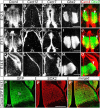
Patterns of Cxcr4, Cxcr7, and Cxcl12 in the early thalamus. AO, Darkfield micrographs of emulsion-dipped coronal sections show the thalamus after in situ hybridization with 35S-labeled probes for Cxcr4 (A, F, K), Cxcl12 (B, G, L), Cxcr7 (C, H, M), and Gbx2 (D, I, N) at E12.5 (A–E), E13.5 (F–J), and E14.5 (K–O). E, J, O, Overlays of false color displays of the adjacent sections shown in A, D; F, I; and K, N. A, E, F, J, K, O, Cxcr4 is expressed in the progenitor domain along the midline and in the Gbx2+ differentiation area of the thalamus. Arrowheads in E, J, and O point to Cxcr4-expressing lateral thalamic nuclei. B, G, L, Cxcl12 is present in the meninges covering the thalamus. C, H, M, Cxcr7 is expressed in the progenitor domain along the midline. A, C, F, H, Arrows point to the zona limitans intrathalamica for orientation. PP″, Confocal images show immunostained GFP and SOX2 in a coronal section of an E14.5 Cxcr4-GFP reporter mouse. 3V, Third ventricle; Hi, hippocampal anlage; Th, thalamus. Scale bars: D, I, N, P′, 500 μm.
Figure 2.
Figure 2.
Transient Cxcr4 expression in the thalamic mantle zone. A, B, D, Dual in situ hybridization for Cxcr4/Sox2 (A, B) and Cxcr4/Gbx2 (D) at E13.5 (A) and E14.5 (B, D). A, B, D, Bright-field views and A′, B′, D′ are dark-field views of the same specimens. A, B, Horizontal and D is a coronal section. Cxcr4 was detected with a 35S-labeled probe (white signal in A′, B′, D′); Sox2 and Gbx2 were detected with digoxigenin (DIG)-labeled probes (A, B, D, blue signal). Cxcr4 is expressed in the Sox2+ and Gbx2+ thalamic mantle zone. C, The H&E-stained coronal section shows the diencephalon, the box corresponds to the thalamic area shown in D. E–G, Boxes in the H&E-stained coronal sections correspond to the thalamic area shown in HP. Images show the thalamus at rostral (top), mid (middle), and caudal levels (bottom). HP, Dark-field micrographs of emulsion-dipped coronal sections after in situ hybridization with 35S-labeled probes for Cxcr4 (HJ), Cxcl12 (KM), and Cxcr7 (NP). 3V, Third ventricle; IC, internal capsule; dLG, dorsal lateral geniculate; GE, ganglionic eminence; Hi, hippocampus; HTh, hypothalamus; Th, thalamus. Scale bars: A, B, D, 200 μm; C, H–J, 500 μm; E–G, 1 mm.
Figure 3.
Figure 3.
CXCL12 activates CXCR4 receptors in the developing thalamus but is dispensable for thalamus formation. AE, Confocal images show immunohistochemistry for inactive/nonphosphorylated CXCR4 (CXCR4inact; A, B, D, E) and total CXCR4 (CXCR4; C) in coronal thalamus sections. A, Overlay of immunostained GFP, CXCR4inact, and DAPI in an E13.5 Cxcr4-GFP reporter mouse shows CXCR4 in the thalamocortical projection (arrowheads). Note that the lateral thalamus (asterisk) contains only sparse CXCR4inact signal despite being Cxcr4-GFP+. B, C, The lateral thalamus (asterisk) shows scant signal for CXCR4inact (B) but strong signal for CXCR4 (C). Sections were counterstained with DAPI. D, E, Immunohistochemistry for CXCR4inact in an E14.5 Cxcl12+/− (control; D) and a Cxcl12−/−littermate (E) demonstrates that CXCR4inact increases in the lateral thalamus (asterisks) in the absence of CXCL12. F, G, Dark-field micrographs of emulsion-dipped coronal sections through the thalamus of an E14.5 Cxcl12+/+ and a Cxcl12−/− littermate after in situ hybridization with a 35S-labeled probe for Sox2. H, Matching thalamus sections, cut at five rostrocaudal sectional levels in E14.5 control (n = 3) and Cxcl12−/− littermates (n = 3), were hybridized for Gbx2 (sectional levels 1, 3, and 5) and Sox2 (sectional levels 2 and 4) as thalamus markers. The marker+ area was determined in micrographs of emulsion-dipped sections after setting a threshold as shown in the insets to the right (signal above threshold appears black). I, J, Confocal images show SOX2-immunoreactivity in the thalamus of an E16.5 Cxcl12+/+ (I) and a Cxcl12−/− littermate (J). K, The thalamus area was determined based on SOX2-immunoreactivity at a rostral and at a caudal sectional level in E16.5 control and Cxcl12−/− littermates (n = 5 each). L, M, X-ray autoradiograms show Slc17a6 (Vglut2) mRNA in hybridized coronal head sections of an E18.5 Cxcl12+/+ (L) and a Cxcl12−/− littermate (M). N, The Slc17a6 mRNA-positive area corresponding to thalamus and epithalamus was determined in x-ray autoradiograms at a rostral and at a caudal sectional level in E18.5 Cxcl12+/+ and Cxcl12−/− littermates (n = 4 each). H, K, N, The thalamus area is not altered in Cxcl12−/− mice (data are mean + SEM). Th, Thalamus. Scale bars: A, C, E, G, I, 200 μm; L, 500 μm.
Figure 4.
Figure 4.
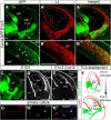
Cxcr4-GFP identifies TCAs. AB″, Confocal images show immunostained GFP and the axonal marker L1 in an E14.5 Cxcr4-GFP reporter mouse. Cxcr4-GFP labels TCAs identified by L1. Presumptive GFP+ interneurons (B, arrowheads) are L1-negative. C, Confocal image of a Cxcr4-GFP reporter mouse shows that GFP+ TCAs have traversed the internal capsule (IC) and reached the cortical SVZ at E13.5. D, E, Dark-field micrographs of emulsion-dipped coronal sections at E13.5 (D) and E14.5 (E) after in situ hybridization with a 35S-labeled probe for Cxcl12. Cxcl12 is expressed in the cortical SVZ, subplate (SP) and meninges (Mn) covering thalamus and cortex. F, Schematic summarizing patterns of Cxcr4-GFP and Cxcl12 mRNA at E13.5 and E14.5. Arrows indicate putative cortical and arrowheads putative meningeal stimulation sites of Cxcr4+ TCAs and thalamic neurons, respectively. G, Dissociated thalamic neurons were immunostained for SOX2 and CXCR4 and counterstained with DAPI. Note CXCR4 in the neurite and the growth-cone. 3V, Third ventricle; Ctx, cortex; GE, ganglionic eminence; Hi, hippocampal anlage; latV, lateral ventricle; Th, thalamus; VZ, ventricular zone. Scale bars: A′, B′, CE, 200 μm; G″20 μm.
Figure 5.
Figure 5.
Deletion of Cxcl12 attenuates intracortical TCA growth. A–H, Thalamic DiI labeling at E14.5 (A, B, E, F) and E16.5 (C, D, G, H) shown in coronal sections at a rostral and a mid-sectional level in control (A–D) and Cxcl12−/− embryos (E–H). Arrowheads point to positions of the most advanced DiI-labeled TCAs. I, J, Mean and SEM of relative intracortical length of DiI-labeled TCAs in E14.5 (I) and E16.5 (J) control embryos and Cxcl12−/− littermates at a rostral and mid-sectional level. K–P, Confocal images show immunostained GFP in control (K–M) and Cxcl12−/− (N–P) E14.5 Cxcr4-GFP reporter mice at rostral (K, N), mid (L, O), and caudal sectional planes (M, P). Arrowheads point to positions of the most advanced GFP+ TCAs. Q, Mean and SEM of relative intracortical length of Cxcr4-GFP+ TCAs in control and Cxcl12−/− E14.5 Cxcr4-GFP reporter mice at rostral, mid, and caudal sectional planes. Statistics in I, J, Q: §§§p < 0.001, §§p < 0.01 for genotype (two-way ANOVA). **p < 0.01, *p < 0.05 (Bonferroni's post hoc test). Numbers of analyzed embryos are indicated. GE, Ganglionic eminence; Hi, hippocampal anlage; latV, lateral ventricle. Scale bars: H, M, 200 μm.
Figure 6.
Figure 6.
A CXCL12/CXCR4 pathway accelerates intracortical TCA growth. A, B, Dark-field views of the cerebral cortex at E14.5 after in situ hybridization with a 35S-labeled probe for Cxcl12. Comparison of a control (A) and a CXCL12-transgenic littermate overexpressing CXCL12 under the Cxcl12 promoter (B) reveals increased Cxcl12 mRNA expression in the transgenic animal. C, D, Confocal images show a Cxcr4-GFP transgenic control (C) and a Cxcr4-GFP;CXCL12-RFP transgenic littermate at E15.5 (D). Immunostained NF is white in C and D, and red in C′, C″, D′, and D″. GFP is green in C′, C″, D′, and D″. DAPI is shown in blue in C, D, C″, and D″. Arrowheads point to the location of the most advanced stained TCAs; asterisks point to TCAs crossing the CP prematurely (D, D′). E, Mean and SEM of relative intracortical length of Cxcr4-GFP+ TCAs in Cxcr4-GFP and Cxcr4-GFP;CXCL12-RFP E15.5 mice in seven sectional planes along the rostrocaudal axis. F, Scheme illustrating measurement of intracortical TCA length. G, Scheme illustrating experimental procedure of explant culturing. Thalamus explants were prepared from E13.5 Cxcr4-GFP mice and cultured for 2 d in medium supplemented with vehicle (control), 20 nm CXCL12, 40 nm CXCL12, or 6 μm CXCR4 antagonist AMD3100. In each explant, length (l) of the 20 longest axons was measured, normalized to control and expressed as mean + SEM. HK, Representative Cxcr4-GFP+ thalamus explants receiving vehicle (H), 40 nm CXCL12 (I), AMD3100 (J), and 40 nm CXCL12 + AMD3100 (K). L, CXCL12 dose-dependently stimulates axonal growth in thalamus explants (for each group, the number of analyzed explants is given in the corresponding bar). Statistics, E: §§§p < 0.001 for genotype (two-way ANOVA), *p < 0.05 (Bonferroni's post hoc test); L: ***p < 0.001, **p < 0.01 (one-way ANOVA and Tukey's post hoc test). Data are mean + SEM. Ctx, Cortex; GE, ganglionic eminence; Hi, hippocampal anlage; latV, lateral ventricle; Mn, meninges; Th, thalamus. Scale bars: A, C (inset), C′, C″, D (inset), H, 200 μm.
Figure 7.
Figure 7.
IPC-derived CXCL12 ensures efficient intracortical TCA growth and interneuron dispersion. A, B, Dark-field micrographs of emulsion-dipped E14.5 coronal brain sections after in situ hybridization with a 35S-labeled probe corresponding to exon 2 of the Cxcl12 gene (exon2 is loxP-flanked in Cxcl12LoxP mice). Cxcl12 is expressed in the cortical SVZ of a Tbr2Cre/+;Cxcl12+/+ (control, A) but not in the SVZ of a Tbr2Cre/+;Cxcl12LoxP/− mouse (Cxcl12cKO, B). Cxcl12 expression is intact in the meninges (Mn) of the Cxcl12cKO. C, D, Confocal images of immunostained GFP in the cortex at mid-rostrocaudal sectional level in E14.5 Cxcl12cKO and control mice carrying the Cxcr4-GFP reporter. Arrowheads pointing to the most advanced GFP+ TCAs demonstrate that GFP+ TCAs are more advanced in the control (C) than in the Cxcl12cKO (D). E, Mean and SEM of relative intracortical length of Cxcr4-GFP+ TCAs in control (n = 8) and Cxcl12cKO embryos (n = 7). F, G, Schemes illustrating the TCA phenotype in the Cxcl12cKO. H, I, Dark-field micrographs of emulsion-dipped coronal sections through the E14.5 cortex after in situ hybridization with a 35S-labeled probe for Lhx6. Fewer Lhx6+ interneurons populate the SVZ (arrows) in the Cxcl12cKO (I) than in the control (H). Note Lhx6+ interneurons are absent in the dorsomedial SVZ in the Cxcl12cKO (I, bracket). J, K, Confocal images of immunostained CXCR4 (white) and DAPI (blue) in coronal cortical sections of E14.5 control (J) and Cxcl12cKO mice (K). Cortical bins are shown in K. There are fewer CXCR4+ interneurons in the SVZ (bins 5–8) and more CXCR4+ interneurons in upper cortical layers (bins 1–3) of the Cxcl12cKO (K) than in the corresponding areas of the control (J). More CXCR4+ interneurons exhibit oblique orientation in the Cxcl12cKO (K, arrowheads) than in the control, indicating abnormal, surface-directed migration of these cells. L, Distribution of CXCR4+ cells in the cortex of E14.5 control (n = 8) and Cxcl12cKO mice (n = 7). Data are mean + SEM of CXCR4+ cells per area in percentage of all CXCR4+ cells. M, Scheme illustrating the interneuron phenotype in the Cxcl12cKO. Statistics: §§§p < 0.0001 for genotype (E) and bin/genotype interaction (L; two-way ANOVA). ***p < 0.001, **p < 0.01, *p < 0.05 (Bonferroni's post hoc test compared with control); n numbers as indicated. GE, ganglionic eminence; latV, lateral ventricle; Mn, meninges; SP, subplate; Th, thalamus; VZ, ventricular zone. Scale bars: A, 500 μm; C, H, J, 200 μm.
Figure 8.
Figure 8.
Schematic summarizing CXCL12/CXCR4 signaling for TCA and interneuron progression. A, Cxcl12-expressing structures are shown in red and Cxcr4-expressing structures in green. B, C, Cxcr4-GFP+ TCAs reach the cortical SVZ at E13.5 and traverse most of the cortex between E13.5 and E14.5. D, E, Intracortical growth of Cxcr4-GFP+ TCAs is reduced in Cxcl12-deficient mice (Cxcl12 KO; D) and in conditional mutants lacking CXCL12 in IPCs (Cxcl12 cKO; E). F, Orthotopic CXCL12 overexpression promotes intracortical growth of Cxcr4-GFP+ TCAs and leads to premature CP invasion and abnormal targeting of the MZ. G, CXCR4-positive interneurons migrate in the MZ, subplate, and SVZ. H, In Cxcl12-deficient mice, interneurons accumulate in the CP/SP area. I, After Cxcl12 ablation in IPCs, interneurons leave the SVZ and seek upper cortical layers. Mn, Meninges; SP, subplate; Th, thalamus.

References

    1. Abe P, Mueller W, Schütz D, MacKay F, Thelen M, Zhang P, Stumm R. CXCR7 prevents excessive CXCL12-mediated downregulation of CXCR4 in migrating cortical interneurons. Development. 2014;141:1857–1863. doi: 10.1242/dev.104224. - DOI - PubMed
    1. Arakawa Y, Bito H, Furuyashiki T, Tsuji T, Takemoto-Kimura S, Kimura K, Nozaki K, Hashimoto N, Narumiya S. Control of axon elongation via an SDF-1alpha/Rho/mDia pathway in cultured cerebellar granule neurons. J Cell Biol. 2003;161:381–391. doi: 10.1083/jcb.200210149. - DOI - PMC - PubMed
    1. Arnold SJ, Huang GJ, Cheung AF, Era T, Nishikawa S, Bikoff EK, Molnár Z, Robertson EJ, Groszer M. The T-box transcription factor Eomes/Tbr2 regulates neurogenesis in the cortical subventricular zone. Genes Dev. 2008;22:2479–2484. doi: 10.1101/gad.475408. - DOI - PMC - PubMed
    1. Auladell C, Pérez-Sust P, Supèr H, Soriano E. The early development of thalamocortical and corticothalamic projections in the mouse. Anat Embryol. 2000;201:169–179. doi: 10.1007/PL00008238. - DOI - PubMed
    1. Bhattacharyya BJ, Banisadr G, Jung H, Ren D, Cronshaw DG, Zou Y, Miller RJ. The chemokine stromal cell-derived factor-1 regulates GABAergic inputs to neural progenitors in the postnatal dentate gyrus. J Neurosci. 2008;28:6720–6730. doi: 10.1523/JNEUROSCI.1677-08.2008. - DOI - PMC - PubMed

Publication types

MeSH terms