. 2015 Sep 23;35(38):13160-70.
doi: 10.1523/JNEUROSCI.3123-14.2015.

Postsynaptic Depolarization Enhances GABA Drive to Dorsomedial Hypothalamic Neurons through Somatodendritic Cholecystokinin Release

Affiliations

Postsynaptic Depolarization Enhances GABA Drive to Dorsomedial Hypothalamic Neurons through Somatodendritic Cholecystokinin Release

Karen M Crosby et al. J Neurosci. .

Abstract

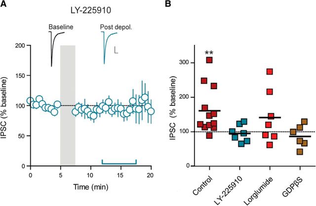
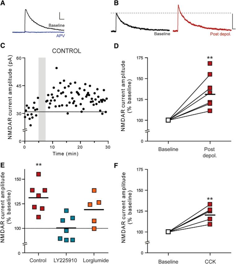
Somatodendritically released peptides alter synaptic function through a variety of mechanisms, including autocrine actions that liberate retrograde transmitters. Cholecystokinin (CCK) is a neuropeptide expressed in neurons in the dorsomedial hypothalamic nucleus (DMH), a region implicated in satiety and stress. There are clear demonstrations that exogenous CCK modulates food intake and neuropeptide expression in the DMH, but there is no information on how endogenous CCK alters synaptic properties. Here, we provide the first report of somatodendritic release of CCK in the brain in male Sprague Dawley rats. CCK is released from DMH neurons in response to repeated postsynaptic depolarizations, and acts in an autocrine fashion on CCK2 receptors to enhance postsynaptic NMDA receptor function and liberate the retrograde transmitter, nitric oxide (NO). NO subsequently acts presynaptically to enhance GABA release through a soluble guanylate cyclase-mediated pathway. These data provide the first demonstration of synaptic actions of somatodendritically released CCK in the hypothalamus and reveal a new form of retrograde plasticity, depolarization-induced potentiation of inhibition. Significance statement: Somatodendritic signaling using endocannabinoids or nitric oxide to alter the efficacy of afferent transmission is well established. Despite early convincing evidence for somatodendritic release of neurohypophysial peptides in the hypothalamus, there is only limited evidence for this mode of release for other peptides. Here, we provide the first evidence for somatodendritic release of the satiety peptide cholecystokinin (CCK) in the brain. We also reveal a new form of synaptic plasticity in which postsynaptic depolarization results in enhancement of inhibition through the somatodendritic release of CCK.

Keywords: GABA; NMDAR; cholecystokinin; dorsomedial hypothalamus; nitric oxide; somatodendritic release.


Figures

Figure 1.
Figure 1.
Repetitive depolarization triggers potentiation of GABA synapses in the DMH. A, Schematic representation of experimental setup (top) and repetitive depolarization protocol (bottom). B, Schematic representation of DMH illustrating the approximate position of recording and stimulating electrodes in the compact zone (CZ) of the DMH. f, Fornix. C, Sample traces of IPSCs before and after repetitive depolarization (top) and a single representative cell (bottom) illustrating potentiation following repetitive depolarization. Traces in all figures are taken from the same cell and averaged for 5 min immediately before (Baseline) and 5–10 min following the end of depolarization (Post depol.). The stimulation artifacts in this and all subsequent figures have been digitally removed for clarity. Calibration: 25 pA, 10 ms. The dotted line represents the average of baseline recording. The shaded area represents the time and duration of depolarization. D, Summary data showing potentiation of GABA synapses following repetitive depolarization (n = 12). The dotted line indicates the baseline, and the solid colored line indicates the duration at which we conducted statistical analysis compared to baseline. Values are mean ± SEM. E, Summary PPR (left) and CV (right) data showing decreases following repetitive depolarization (the black bars in these and subsequent figures indicate the means). F, Summary sIPSC data showing an increase in frequency and no change in amplitude following repetitive depolarization. Inset, Representative sIPSC traces taken before and after repetitive depolarization. Calibration: 25 pA, 0.5 s. *p < 0.05.
Figure 2.
Figure 2.
Postsynaptic vesicular release is required for DPI. A, Sample traces of IPSCs before and after repetitive depolarization (top) and summary data from each cell (bottom; n = 7) showing no potentiation in the presence of the SNAP-25 blocking peptide (50 μm; included in the patch pipette). B, Sample traces of IPSCs before and after repetitive depolarization (top) and summary data from each cell (bottom; n = 8) showing potentiation in the presence of the SNAP-25 scrambled peptide (50 μm; included in the patch pipette). Calibrations: 25 pA, 10 ms. **p < 0.01 versus baseline.
Figure 3.
Figure 3.
CCK-induced activation of CCK2Rs mediates DPI. A, Sample traces of IPSCs before and after repetitive depolarization in the presence of the CCK2R antagonist LY-225910 (1 μm; top) and summary data showing that LY-225910 completely blocks potentiation of GABA synapses following repetitive depolarization (bottom; n = 7). Calibration: 25 pA, 10 ms. B, Summary data showing the percentage change in IPSC amplitude following repetitive depolarization in the following conditions: control (n = 12), LY-225910 (n = 7), lorglumide (CCK1R antagonist; 1 μm; n = 7), and GDPβS (1 mm; in the patch pipette; n = 6). **p < 0.01 versus baseline.
Figure 4.
Figure 4.
Exogenous application of CCK-8S enhances GABA release through a CCK2R- and NO-mediated pathway. A, Sample traces of IPSCs before and during application of CCK-8S (0.1 μm; top) and a single representative cell (bottom) illustrating potentiation in response to CCK-8S application (duration of application indicated by colored bar). Calibration: 25 pA, 10 ms. B, Summary data from each cell showing potentiation of IPSCs with CCK-8S application (n = 12). C, Summary PPR (left) and CV (right) data showing decreases with CCK application. D, Summary sIPSC data showing an increase in frequency and no change in amplitude with CCK application. Inset, Representative sIPSC traces taken before and during CCK-8S application. Calibration: 25 pA, 0.5 s. E, Summary data from each cell showing no change in IPSCs with CCK-8S application in the continuous presence of LY-225910 (CCK2R antagonist; n = 7). F, Summary data from each cell showing no change in IPSCs with CCK-8S application in the continuous presence of l-NAME (NOS inhibitor; n = 7). *p < 0.05; **p < 0.01 versus baseline.
Figure 5.
Figure 5.
NMDAR activation and retrograde NO signaling are necessary for DPI. A, Sample traces of IPSCs before and after repetitive depolarization in the presence of the NO synthase inhibitor l-NAME (200 μm; top) and summary data from each cell showing l-NAME completely blocks potentiation of GABA synapses following repetitive depolarization (bottom; n = 6). B, Sample traces of IPSCs before and after repetitive depolarization in the presence of the sGC inhibitor ODQ (10 μm; top) and summary data from each cell showing ODQ completely blocks potentiation of GABA synapses following repetitive depolarization (bottom; n = 6). C, Sample traces of IPSCs before and after repetitive depolarization in the presence of the NMDAR antagonist APV (50 μm; top) and summary data from each cell showing APV completely blocks potentiation of GABA synapses following repetitive depolarization (bottom; n = 7). Calibrations: 25 pA, 10 ms.
Figure 6.
Figure 6.
Repetitive depolarization triggers potentiation of NMDA currents. A, Sample traces of NMDAR-mediated currents before (black) and during incubation with the NMDAR antagonist APV (50 μm; blue). B, Sample traces of NMDAR-mediated currents before (left) and after repetitive depolarization (right). C, Single representative cell illustrating potentiation in NMDAR-mediated currents following repetitive depolarization. D, Summary data from each cell showing potentiation in NMDAR-mediated currents following repetitive depolarization. E, Summary data from each cell showing percentage change in NMDA current amplitude following repetitive depolarization in the following conditions: control (n = 7), LY-225910 (CCK2R antagonist; n = 7), and lorglumide (CCK1R antagonist; n = 5). F, Summary data from each cell showing that exogenous application of CCK enhances NMDA current amplitude (n = 5). **p < 0.01 versus baseline. Calibrations: 25 pA, 10 ms.
Figure 7.
Figure 7.
Schematic representation of somatodendritic CCK release and subsequent DPI. Postsynaptic depolarization (1) triggers somatodendritic CCK release (2). CCK activates postsynaptic CCK2Rs (3), which increase NMDAR activity (4). NMDAR activation triggers NO production through coupling to neuronal NOS (5), and NO acts as a retrograde signal (6) to enhance GABA release from the presynaptic terminal (7).

References

    1. Abelson JL, Young EA. Hypothalamic-pituitary adrenal response to cholecystokinin-B receptor agonism is resistant to cortisol feedback inhibition. Psychoneuroendocrinology. 2003;28:169–180. doi: 10.1016/S0306-4530(02)00013-6. - DOI - PubMed
    1. Bains JS, Ferguson AV. Nitric oxide regulates NMDA-driven GABAergic inputs to type I neurones of the rat paraventricular nucleus. J Physiol. 1997;499:733–746. doi: 10.1113/jphysiol.1997.sp021965. - DOI - PMC - PubMed
    1. Benquet P, Gee CE, Gerber U. Two distinct signaling pathways upregulate NMDA receptor responses via two distinct metabotropic glutamate receptor subtypes. J Neurosci. 2002;22:9679–9686. - PMC - PubMed
    1. Bi S, Ladenheim EE, Schwartz GJ, Moran TH. A role for NPY overexpression in the dorsomedial hypothalamus in hyperphagia and obesity of OLETF rats. Am J Physiol Regul Integr Comp Physiol. 2001;281:R254–R260. - PubMed
    1. Bi S, Scott KA, Kopin AS, Moran TH. Differential roles for cholecystokinin A receptors in energy balance in rats and mice. Endocrinology. 2004;145:3873–3880. doi: 10.1210/en.2004-0284. - DOI - PubMed

Publication types

MeSH terms

LinkOut - more resources