. 2015 Sep 23;35(38):13206-18.
doi: 10.1523/JNEUROSCI.0193-15.2015.

Aging-Related Hyperexcitability in CA3 Pyramidal Neurons Is Mediated by Enhanced A-Type K+ Channel Function and Expression

Affiliations

Aging-Related Hyperexcitability in CA3 Pyramidal Neurons Is Mediated by Enhanced A-Type K+ Channel Function and Expression

Dina Simkin et al. J Neurosci. .

Abstract

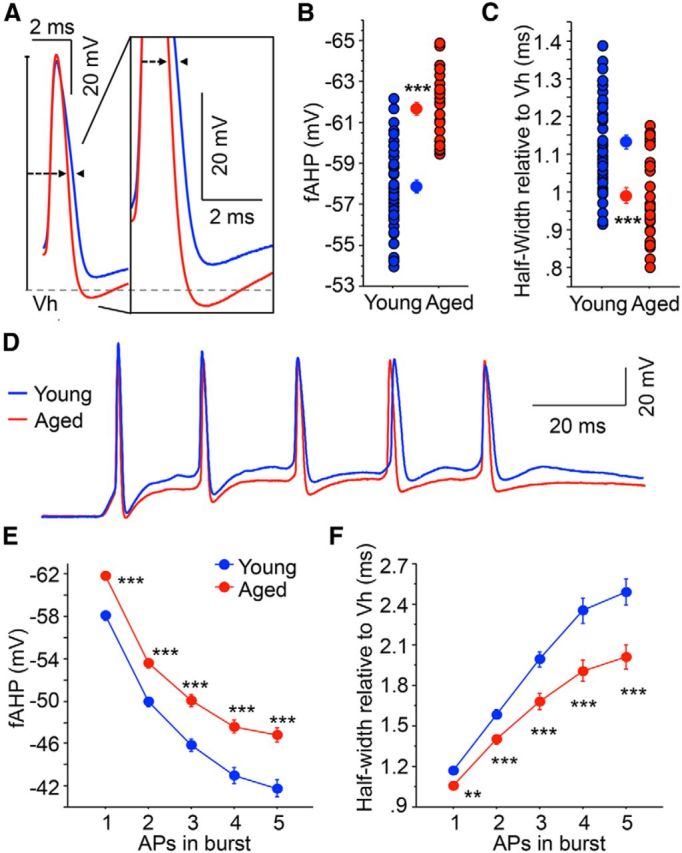
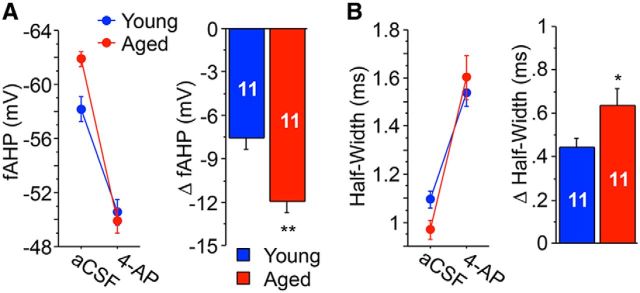
Aging-related impairments in hippocampus-dependent cognition have been attributed to maladaptive changes in the functional properties of pyramidal neurons within the hippocampal subregions. Much evidence has come from work on CA1 pyramidal neurons, with CA3 pyramidal neurons receiving comparatively less attention despite its age-related hyperactivation being postulated to interfere with spatial processing in the hippocampal circuit. Here, we use whole-cell current-clamp to demonstrate that aged rat (29-32 months) CA3 pyramidal neurons fire significantly more action potentials (APs) during theta-burst frequency stimulation and that this is associated with faster AP repolarization (i.e., narrower AP half-widths and enlarged fast afterhyperpolarization). Using a combination of patch-clamp physiology, pharmacology, Western blot analyses, immunohistochemistry, and array tomography, we demonstrate that these faster AP kinetics are mediated by enhanced function and expression of Kv4.2/Kv4.3 A-type K(+) channels, particularly within the perisomatic compartment, of CA3 pyramidal neurons. Thus, our study indicates that inhibition of these A-type K(+) channels can restore the intrinsic excitability properties of aged CA3 pyramidal neurons to a young-like state. Significance statement: Age-related learning deficits have been attributed, in part, to altered hippocampal pyramidal neuronal function with normal aging. Much evidence has come from work on CA1 neurons, with CA3 neurons receiving comparatively less attention despite its age-related hyperactivation being postulated to interfere with spatial processing. Hence, we conducted a series of experiments to identify the cellular mechanisms that underlie the hyperexcitability reported in the CA3 region. Contrary to CA1 neurons, we demonstrate that postburst afterhyperpolarization is not altered with aging and that aged CA3 pyramidal neurons are able to fire significantly more action potentials and that this is associated with faster action potential repolarization through enhanced expression of Kv4.2/Kv4.3 A-type K(+) channels, particularly within the cell bodies of CA3 pyramidal neurons.

Keywords: A-type K+ channels; CA3; Kv4.2/Kv4.3; action potential repolarization; aging; pyramidal neurons.


Figures

Figure 1.
Figure 1.
Medium and slow postburst AHPs are not altered with aging in CA3 pyramidal neurons. A, Schematic of hippocampal slice section illustrating the placement of recording and stimulating electrode in the CA3 region. B, An example of a histochemical section stained with DAB showing morphology of two representative CA3 neurons filled with neurobiotin from patch pipettes during whole-cell patch-clamp experiments. C, Illustrated are examples of postburst AHPs evoked with a 50 Hz train of suprathreshold current injections into young (blue) and aged (red) CA3 pyramidal neurons. Medium and slow postburst AHPs were measured at the peak and 1 s interval after last current pulse, respectively. D, There was no age-related difference in either the medium or slow postburst AHP from recorded CA3 pyramidal neurons. Also, changing the aCSF solution from normal aCSF to aCSF with GABAR blockers had no effect on current injection-elicited AHPs in either young or aged CA3 neurons (data not shown). E, Representative voltage traces of AHPs evoked by 5 suprathreshold synaptic stimuli at 50 Hz before and after the addition of GABAR blockers to the aCSF recorded from young and aged CA3 neurons. F, There were no age-related differences in either peak or 1 s AHP (see Table 2) before and after GABAergic inhibition. C, E, APs are truncated for clarity. Values are mean ± SEM. The number of cells (n) is represented in the middle of each bar for each group.
Figure 2.
Figure 2.
Excitability is enhanced in aged CA3 pyramidal neurons. A, Examples of representative traces from young (blue) and aged (red) CA3 pyramidal neurons are illustrated that demonstrate increased firing (or reduced accommodation) in aged CA3 pyramidal neurons to a 100 ms, 100 Hz train of synaptic stimuli. The stimulus intensity was set at threshold voltage to evoke an AP for the first stimulus in the 100 Hz train. B, Significantly more APs were observed in aged CA3 neurons during the 100 ms, 100 Hz train. C, Examples of representative traces from aged and young CA3 neurons are illustrated that show increased firing evoked during theta burst stimulation. Each 100 Hz burst (as described in A) was delivered 5 times at 5 Hz. D, Repeated-measures ANOVA revealed that aged neurons fired more APs during theta burst stimulation protocol compared with young. There was also a significant interaction of age and burst number (1–5). E, The IPSP or EPSP amplitude was measured as a function of synaptic subthreshold stimulation intensity. The slope of this relationship was determined from each cell. There was no age-related difference in IPSP (left; p = 0.98) or EPSP (right; p = 0.82) input–output slopes; thus, synaptic drive was similar. *p < 0.05 (Fisher's PLSD t test). **p < 0.005 (Fisher's PLSD t test). Values are mean ± SEM. The number of cells (n) is represented in the middle of each bar for each group.
Figure 3.
Figure 3.
Aged CA3 pyramidal neurons exhibit increased fAHP and reduced spike broadening. A, Illustrated are overlays of representative traces from young (blue; n = 43) and aged (red; n = 28) CA3 neurons that show fAHP evoked with single orthodromic AP. fAHP is measured as absolute membrane voltage. AP half-width is measured as the duration of the AP at half of the AP amplitude (relative to holding potential −60 mV). B, The fAHP evoked with a single orthodromic stimulus was significantly enhanced (more hyperpolarized) in aged CA3 neurons than young neurons. C, AP half-width was significantly narrower in aged CA3 neurons than young neurons. D, Overlay of representative traces of young (blue) and aged (red) CA3 neurons after repeated orthodromic stimulations at suprathreshold intensities (50 Hz). E, Significantly larger fAHPs were observed in aged CA3 neurons after 5 synaptically evoked APs at 50 Hz (repeated-measures ANOVA, F(1,276) = 29.58, p < 0.0001). Planned post hoc analysis further revealed that the fAHP was significantly different after each of the 5 synaptically evoked APs at 50 Hz. F, Significantly less spike broadening was observed in aged CA3 neurons after a 50 Hz train of 5 suprathreshold orthodromic APs. Repeated-measures ANOVA revealed not only a decrease in the spike half-width with age but also an age by spike number interaction, suggesting that the rate of change with each subsequent spike is higher in young CA3 neurons. **p < 0.005 (Fisher's PLSD t test). ***p < 0.0005 (Fisher's PLSD t test). Values are mean ± SEM.
Figure 4.
Figure 4.
A-type K+ channel blocker 4-AP produces a larger magnitude reduction in fAHP and increase in half-width in aged CA3 neurons than young. A, The fAHP was recorded after a single orthodromically induced AP before and after the addition of 2 mm 4-AP to the aCSF bathing solution. 4-AP incubation caused a larger magnitude reduction in fAHP in aged CA3 neurons compared with young CA3 neurons. B, 4-AP application also caused a larger magnitude increase in AP half-width in aged neurons compared with young neurons. *p < 0.05 (Fisher's PLSD t test), **p < 0.005 (Fisher's PLSD t test). Values are represented as mean ± SEM. The number of cells (n) is represented in the middle of each bar for each group.
Figure 5.
Figure 5.
Inhibition of Kv4.2/Kv4.3 K+ channels with PaTx reverses aging-related alterations in fAHP and AP half-width. A, Representative traces of single orthodromically elicited APs from aged (red) and young (blue) CA3 neurons with (dashed line) and without (solid line) 1 μm PaTx in recording pipette (from AP threshold). PaTx diffusion into the cell significantly reduced the fAHP of aged CA3 neurons with no significant effect in young after a single orthodromically induced AP (B) and after 5 synaptically evoked AP at 50 Hz (C). PaTx also enhanced AP half-widths in aged CA3 neurons after single (D) or 5 sequential (E) synaptically evoked APs to durations similar to young CA3 neurons while having no effect on young half-widths (see Table 3). *p < 0.05 (Fisher's PLSD t test). **p < 0.005 (Fisher's PLSD t test). ***p < 0.0005 (Fisher's PLSD t test). Values are mean ± SEM. The number of cells (n) is represented in the figure legend.
Figure 6.
Figure 6.
Faster AP kinetics in aged CA3 neurons are not mediated by BK channels. A, Addition of BK channel antagonist, paxilline, to the aCSF bathing solution significantly reduced the fAHP of a single orthodromic-elicited AP of both young and aged neurons. B, Paxilline also increased the half-width of both young and aged CA3 neurons by similar magnitude after a single orthodromically evoked AP. Five synaptically evoked APs before and after the addition of paxilline induced a reduction in fAHP (C) and an increase in half-width (D) for both young and aged CA3 neurons. The magnitude of fAHP (E) and half-width (F) change was calculated for each AP spike. Repeated-measures ANOVA did not reveal an age effect for ΔfAHP or Δhalf-width measures (ΔfAHP: p = 0.37, Δhalf-width: p = 0.74, n = 8 young and n = 7 aged). Values are mean ± SEM. A, C, The number of cells (n) is represented in the middle of each bar for each group.
Figure 7.
Figure 7.
A-type Kv4 channel expression is enhanced in the membrane fraction of aged CA3. A, Western blot analysis of CA3 hippocampal tissue from young (blue) and aged (red) rats using BK, Kv4.2, and Kv4.3 antibodies. Loading control total protein stained with Sypro (large blot on the right). B, The optical density values for BK, Kv4.2, and Kv4.3 (relative to total membrane protein as a loading control) were normalized to the young rat CA3 tissue optical densities for each Western blot. Whereas BK channel expression was similar between young adult and aged rats, expression of both Kv4.2 and Kv4.3 was increased. Numbers report p values for the independent-samples t tests. *p = 0.02 (Fisher's PLSD t test).
Figure 8.
Figure 8.
Perisomatic redistribution of A-type K+ channels in aged hippocampal CA3 region. A, Monochrome images of free-floating brain sections immunostained for Kv4.2 channels from a young adult (top) and an aged (bottom) rat. Pink arrows indicate approximate borders of hippocampal region CA3. Unfilled arrows indicate approximate borders of CA3b, which was targeted in the patch-clamp experiments. Inset, Triple-immunofluorescence image of aged pyramidal neuron from which patch-clamp data were obtained (green), with Kv4.2 immunofluorescence (red) and DAPI (blue). Scale bar, 250 μm. B, Monochrome images at lower magnification (left) and higher magnification (right) of Kv4.2 immunoreactivity in hippocampal slices used in patch-clamp experiments from a young adult (top) and an aged (bottom) rat. Purple arrows in this and all other panels indicate the approximate borders between the perisomatic region (SP and SL) and stratum oriens (so) and SR (sr). Scale bars: left, 150 μm; right, 50 μm. C, D, Monochrome array tomography immunofluorescence images at low (C) and higher (D) magnification from brain slices used in patch-clamp experiments obtained from young (C, D, left panel) and aged (C, D, right panel) rats. Scale bars: C, 75 μm; D, 25 μm. E–H, Same as A–D, but for Kv4.3 immunoreactivity. I, Ratio of perisomatic-to-dendritic immunosignal in CA3 from brain slice immunofluorescence experiments (circles) and array tomography immunofluorescence experiments (triangles) in young adult (blue) and aged (red) rats normalized to young adult rat values. *p < 0.05, significant difference (unpaired t test). J, Same as I, but for Kv4.3 immunoreactivity. I, *p < 0.05, significant difference (unpaired t test). I–J, Lines connect data from the paired cohorts. Group means are presented as ± SEM.

Similar articles

Cited by

References

    1. Amaral DG, Witter MP. The 3-dimensional organization of the hippocampal formation: a review of anatomical data. Neuroscience. 1989;31:571–591. doi: 10.1016/0306-4522(89)90424-7. - DOI - PubMed
    1. Andrásfalvy BK, Makara JK, Johnston D, Magee JC. Altered synaptic and non-synaptic properties of CA1 pyramidal neurons in Kv4.2 knockout mice. J Physiol. 2008;586:3881–3892. doi: 10.1113/jphysiol.2008.154336. - DOI - PMC - PubMed
    1. Aronica E, Boer K, Doorn KJ, Zurolo E, Spliet WG, van Rijen PC, Baayen JC, Gorter JA, Jeromin A. Expression and localization of voltage dependent potassium channel Kv4.2 in epilepsy associated focal lesions. Neurobiol Dis. 2009;36:81–95. doi: 10.1016/j.nbd.2009.06.016. - DOI - PubMed
    1. Bakker A, Krauss GL, Albert MS, Speck CL, Jones LR, Stark CE, Yassa MA, Bassett SS, Shelton AL, Gallagher M. Reduction of hippocampal hyperactivity improves cognition in amnestic mild cognitive impairment. Neuron. 2012;74:467–474. doi: 10.1016/j.neuron.2012.03.023. - DOI - PMC - PubMed
    1. Burke SN, Barnes CA. Neural plasticity in the ageing brain. Nat Rev. 2006;7:30–40. doi: 10.1038/nrn1809. - DOI - PubMed

Publication types

MeSH terms

Substances