. 2015 Nov 1;195(9):4085-95.
doi: 10.4049/jimmunol.1402455. Epub 2015 Sep 23.

Definition of Proteasomal Peptide Splicing Rules for High-Efficiency Spliced Peptide Presentation by MHC Class I Molecules

Affiliations

Definition of Proteasomal Peptide Splicing Rules for High-Efficiency Spliced Peptide Presentation by MHC Class I Molecules

Celia R Berkers et al. J Immunol. .

Abstract

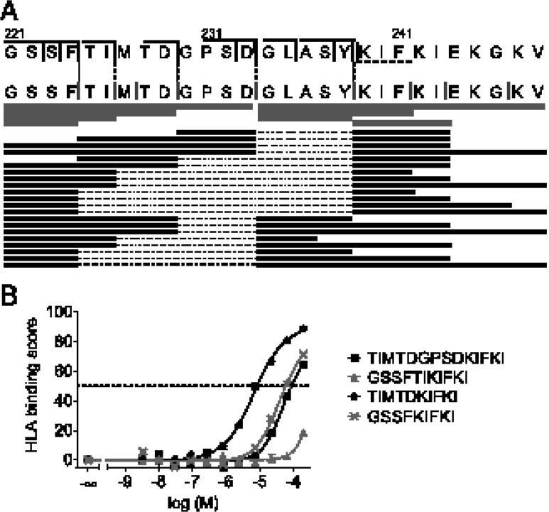
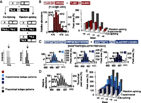
Peptide splicing, in which two distant parts of a protein are excised and then ligated to form a novel peptide, can generate unique MHC class I-restricted responses. Because these peptides are not genetically encoded and the rules behind proteasomal splicing are unknown, it is difficult to predict these spliced Ags. In the current study, small libraries of short peptides were used to identify amino acid sequences that affect the efficiency of this transpeptidation process. We observed that splicing does not occur at random, neither in terms of the amino acid sequences nor through random splicing of peptides from different sources. In contrast, splicing followed distinct rules that we deduced and validated both in vitro and in cells. Peptide ligation was quantified using a model peptide and demonstrated to occur with up to 30% ligation efficiency in vitro, provided that optimal structural requirements for ligation were met by both ligating partners. In addition, many splicing products could be formed from a single protein. Our splicing rules will facilitate prediction and detection of new spliced Ags to expand the peptidome presented by MHC class I Ags.


Figures

FIGURE 1.
FIGURE 1.
Splicing rules predict splicing-prone peptide sequences that can participate in highly efficient ligation reactions. (A) Summary of the splicing rules determined for the C-terminal fragment (gray circles, P1′, P2′, and P3′ residues) and the N-terminal precursor (black circles, P1, P2 residues). Sx′/Sx = proteasomal substrate binding pockets. Φ = hydrophobic residue. (B) Relative ligation efficiencies of different C-terminal ligation fragments measured in fluorescence polarization assays, indicating splicing preferences for the P1′, P2′, and P3′ positions. Efficiencies were normalized to the efficiency of [YLDW][KLPSV] formation, which was included in all experiments. Values are averaged over at least two independent experiments; error bars represent SD. (C and D) Relative maximum ligation efficiencies of different N-terminal ligation precursors measured in LC-MS assays, indicating splicing preferences for the P1 and P2 positions. Maximum efficiencies are averages of the two highest efficiencies as measured in time-course experiments and normalized to the maximum efficiency of [YLGL][RLPSV] formation, which was included in all experiments. Error bars represent SD. (E) Ligation efficiencies of different ligation products resulting from digestion of the optimized splicing precursor YLGD-SY-RLPSV, measured at various time points and precursor peptide concentration.
FIGURE 2.
FIGURE 2.
Splicing rules facilitate the prediction of spliced epitopes from a long polypeptide. (A) Top panel, Peptide 221-248/C223S (N1.1) derived from the neuraminidase 1 protein of Influenza A virus A/Puerto Rico/8/34, showing several predicted N-terminal precursor sequences (black solid vertical lines) and one predicted C-terminal ligation motif (black dotted vertical lines). Middle panel, Actual cleavage sites in peptide 221-248 (gray vertical lines) and detected hydrolysis products (gray horizontal bars), measured by LC-MS. Bottom panel, Overview of ligation products measured in digestion mixtures of N1.1 by LC-MS, indicated by black horizontal bars, where the excised fragment is indicated by a dotted line. (B) HLA-A*0201 binding curves of selected splicing products originating from N1.1.
FIGURE 3.
FIGURE 3.
Splicing of long peptides predominantly occurs via cis-splicing. (A) Splicing of a mixture of 15N-Gly–labeled (indicated in bold) and unlabeled precursor peptides through cis-splicing or through random splicing results in differently labeled spliced products and therefore in different isotope patterns that can be distinguished by LC-MS. (B) Left panel, Mass spectrum (black trace) and the corresponding experimental isotope pattern (red bars) of [YLGD][KLGSV] resulting from the digestion of a mixture of 15N-labeled and unlabeled precursor YLGD-SY-KLGSV. Right panel, Comparison of the experimental isotope pattern with theoretical isotope patterns for cis- and random splicing. (C) Mass spectra (black traces) and the corresponding experimental isotope pattern (blue bars) of [GSSFTIMTD][GLASYKIFKIEKGKV], resulting from the digestion of a mixture of 15N-Gly–labeled (indicated in bold) and unlabeled precursor GSSFTIMTD-GPSD-GLASYKIFKIEKGKV for different time periods. (D) Mass spectrum (black trace) of a 1:1 mixture of single-isotope incubation samples, in which the labeled and 15N2-labeled splicing products are present in equal amounts, compared with the theoretical isotope pattern for cis-splicing (gray bars). (E) Comparison of the experimental isotope patterns (blue bars) determined in (C) with theoretical isotope patterns for cis- and random splicing.
FIGURE 4.
FIGURE 4.
Splicing rules predict efficient splicing reactions in cells. (A) Ion traces and estimated amounts of the test peptide SGYSGIFSVEGK (10 fmol on column), the spliced peptides [YLGN][RLPSV] and [YLGD][RLPSV], and the HLA-A*0201 binding peptide YLWGRPLSV, as eluted from the cell surface of JY or MelJuso-A2 cells. (B) MS/MS spectrum of peptide [YLGD][RLPSV] as eluted from the cell surface of JY cells.

References

    1. Neefjes J., Jongsma M. L., Paul P., Bakke O. 2011. Towards a systems understanding of MHC class I and MHC class II antigen presentation. Nat. Rev. Immunol. 11: 823–836. - PubMed
    1. Sijts E. J., Kloetzel P. M. 2011. The role of the proteasome in the generation of MHC class I ligands and immune responses. Cell. Mol. Life Sci. 68: 1491–1502. - PMC - PubMed
    1. Vigneron N., Van den Eynde B. J. 2012. Proteasome subtypes and the processing of tumor antigens: increasing antigenic diversity. Curr. Opin. Immunol. 24: 84–91. - PubMed
    1. Basler M., Kirk C. J., Groettrup M. 2013. The immunoproteasome in antigen processing and other immunological functions. Curr. Opin. Immunol. 25: 74–80. - PubMed
    1. Vigneron N., Stroobant V., Chapiro J., Ooms A., Degiovanni G., Morel S., van der Bruggen P., Boon T., Van den Eynde B. J. 2004. An antigenic peptide produced by peptide splicing in the proteasome. Science 304: 587–590. - PubMed

Publication types

MeSH terms

Substances

LinkOut - more resources