

Xanthogranulomatous cholecystitis: Difficulty in differentiating from gallbladder cancer
- PMID: 26401081
- PMCID: PMC4572797
- DOI: 10.3748/wjg.v21.i35.10166
Xanthogranulomatous cholecystitis: Difficulty in differentiating from gallbladder cancer
Abstract
Aim: To compare cases of xanthogranulomatous cholecystitis (XGC) and advanced gallbladder cancer and discuss the differential diagnoses and surgical options.
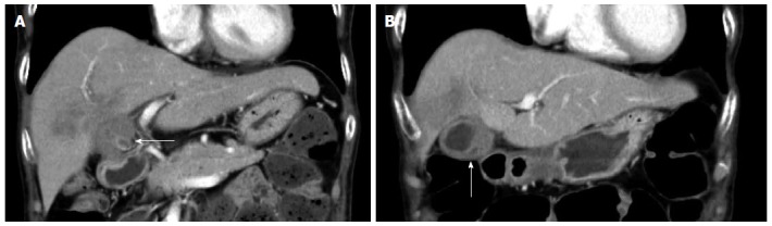
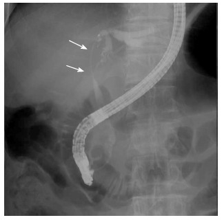
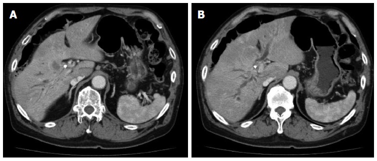
Methods: From April 2000 to December 2013, 6 XGC patients received extended surgical resections. During the same period, 16 patients were proven to have gallbladder (GB) cancer, according to extended surgical resection. Subjects chosen for analysis in this study were restricted to cases of XGC with indistinct borders with the liver as it is often difficult to distinguish these patients from those with advanced GB cancer. We compared the clinical features and computed tomography findings between XGC and advanced GB cancer. The following clinical features were retrospectively assessed: age, gender, symptoms, and tumor markers. As albumin and the neutrophil/lymphocyte ratio (NLR) are prognostic in several cancers, we compared serum albumin levels and the NLR between the two groups. The computerized tomography findings were used to compare the two diseases, determine the coexistence of gallstones, the pattern of GB thickening (focal or diffuse), the presence of a hypoattenuated intramural nodule, and continuity of the mucosal line.
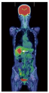
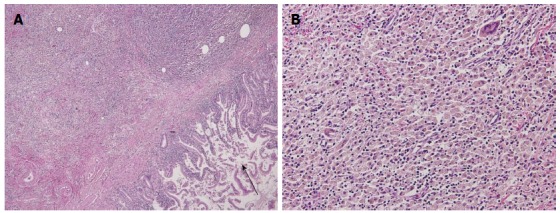
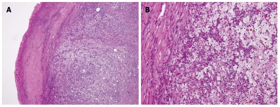
Results: Based on the preoperative image findings, we suspected GB carcinoma in all cases including XGC in this series. In addition, by pathological examination, we found that the group of patients with XGC developed inflammatory disease after surgery. Patients with XGC tended to have abdominal pain (4/6, 67%). However, there was no significant difference in clinical symptoms, including fever, between the two groups. Serum albumin and NLR were also similar in the two groups. Serum tumor markers, such as carcinoembryonic antigen (CEA) and carbohydrate antigen 19-9 (CA19-9), tended to increase in patients with GB cancer. However, no significant differences in tumor markers were identified. On the other hand, gallstones were more frequently observed in patients with XGC (5/6, 83%) than in patients with GB cancer (4/16, 33%) (P = 0.0116). A hypoattenuated intramural nodule was found in 3 patients with XGC (3/6, 50%), but in only 1 patient with GB cancer (1/16, 6%) (P = 0.0024). The GB thickness, continuous mucosal line, and bile duct dilatation showed no significant differences between XGC and GB cancer.
Conclusion: Although XGC is often difficult to differentiate from GB carcinoma, it is possible to obtain an accurate diagnosis by careful intraoperative gross observation, and several intraoperative frozen sections.
Keywords: Advanced gallbladder cancer; Differential diagnosis; Xanthogranulomatous cholecystitis.
Figures
References
-
- Benbow EW. Xanthogranulomatous cholecystitis. Br J Surg. 1990;77:255–256. - PubMed
-
- Spinelli A, Schumacher G, Pascher A, Lopez-Hanninen E, Al-Abadi H, Benckert C, Sauer IM, Pratschke J, Neumann UP, Jonas S, et al. Extended surgical resection for xanthogranulomatous cholecystitis mimicking advanced gallbladder carcinoma: A case report and review of literature. World J Gastroenterol. 2006;12:2293–2296. - PMC - PubMed
-
- Yang T, Zhang BH, Zhang J, Zhang YJ, Jiang XQ, Wu MC. Surgical treatment of xanthogranulomatous cholecystitis: experience in 33 cases. Hepatobiliary Pancreat Dis Int. 2007;6:504–508. - PubMed
Publication types
MeSH terms
Supplementary concepts
LinkOut - more resources
Full Text Sources
Other Literature Sources
Medical
Research Materials
Miscellaneous
