. 2014 Apr 16:1:4.
doi: 10.1186/2052-6687-1-4. eCollection 2014.

A novel mutation in TTC8 is associated with progressive retinal atrophy in the golden retriever

Affiliations

A novel mutation in TTC8 is associated with progressive retinal atrophy in the golden retriever

Louise M Downs et al. Canine Genet Epidemiol. .

Abstract

Background: Generalized progressive retinal atrophy (PRA) is a group of inherited eye diseases characterised by progressive retinal degeneration that ultimately leads to blindness in dogs. To date, more than 20 different mutations causing canine-PRA have been described and several breeds including the Golden Retriever are affected by more than one form of PRA. Genetically distinct forms of PRA may have different clinical characteristics such as rate of progression and age of onset. However, in many instances the phenotype of different forms of PRA cannot be distinguished at the basic clinical level achieved during routine ophthalmoscopic examination. Mutations in two distinct genes have been reported to cause PRA in Golden Retrievers (prcd-PRA and GR_PRA1), but for approximately 39% of cases in this breed the causal mutation remains unknown.

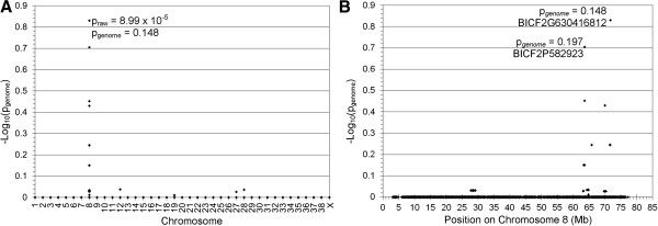
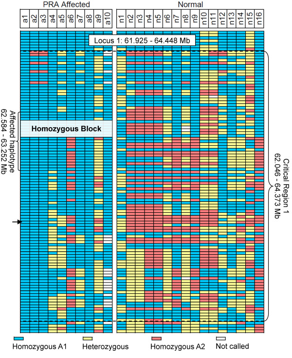
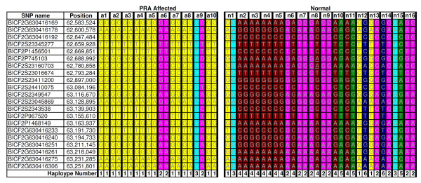
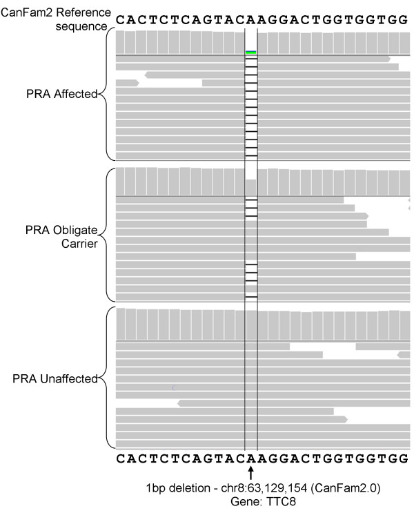
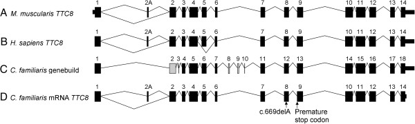
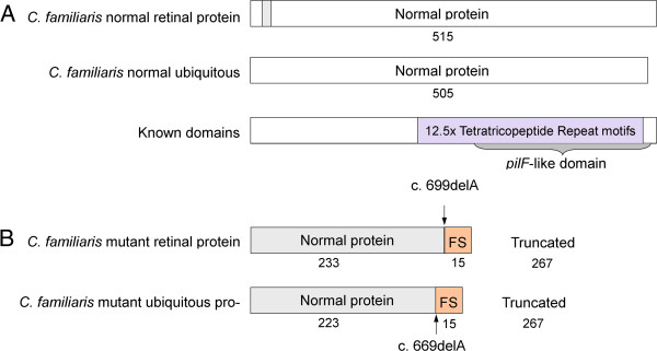
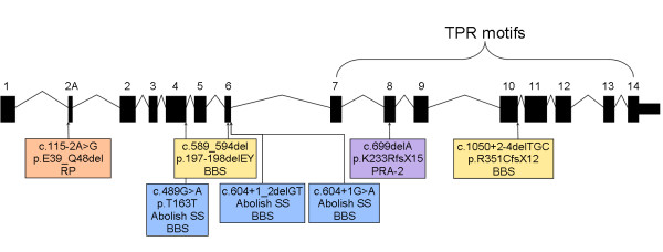
Results: A genome-wide association study of 10 PRA cases and 16 controls identified an association on chromosome 8 not previously associated with PRA (praw = 1.30×10(-6) and corrected with 100,000 permutations, pgenome = 0.148). Using haplotype analysis we defined a 737 kb critical region containing 6 genes. Two of the genes (TTC8 and SPATA7) have been associated with Retinitis Pigmentosa (RP) in humans. Using targeted next generation sequencing a single nucleotide deletion was identified in exon 8 of the TTC8 gene of affected Golden Retrievers. The frame shift mutation was predicted to cause a premature termination codon. In a larger cohort, this mutation, TTC8 c.669delA, segregates correctly in 22 out of 29 cases tested (75.9%). Of the PRA controls none are homozygous for the mutation, only 3.5% carry the mutation and 96.5% are homozygous wildtype.

Conclusions: Our results show that PRA is genetically heterogeneous in one of the world's numerically largest breeds, the Golden Retriever, and is caused by multiple, distinct mutations. Here we discuss the mutation that causes a form of PRA, that we have termed PRA2, that accounts for approximately 30% of PRA cases in the breed. The genetic explanation for approximately 9% of cases remains to be identified. PRA2 is a naturally occurring animal model for Retinitis Pigmentosa, and potentially Bardet-Biedl Syndrome.


Figures

Figure 1
Figure 1
Genome-wide association mapping of PRA in Golden Retrievers. -Log10 of p-values after correction for multiple testing and population stratification with 100,000 permutations and IBS clustering, respectively. (A) -Log10 plot of genome-wide association results show a single, albeit not statistically significant, signal on CFA8 (praw = 8.99 × 10-5, pgenome = 0.148). The most significant of the raw and permuted values are indicated. (B) The associated SNPs on CFA8 form two distinct signals with the most associated SNPs at 63.614 Mb and 71.732 Mb. Permuted values at these loci are indicated.
Figure 2
Figure 2
Critical region definition using homozygosity analysis. SNP genotypes for 10 PRA cases and 16 PRA controls, over one of the two regions identified during the GWA study. The most associated SNP in the region, BICF2P582923 (Marker 1) at 63.614 is indicated with an arrow. All 10 cases, as well as 5 of the controls, share a 437 kb homozygous block, while 8 cases and none of the controls share a larger homozygous region (Affected haplotype) upstream of Marker 1.
Figure 3
Figure 3
Fine mapping using haplotype analysis. SNP genotypes for 10 PRA cases and 16 PRA controls, over the 668 kb “Affected haplotype” identified during the GWA study. Inferred phasing of the 21 SNP markers revealed six unique haplotypes. Haplotype number 1 (yellow) is homozygous in 8/10 cases, but none of the controls.
Figure 4
Figure 4
IGV display of the 1-bp deletion in TTC8 (c.669delA). Each of the three samples (PRA-affected, obligate carrier and control) viewed in IGV are represented by two panels. The upper panel is a histogram where the height of each column is representative of the read depth at that location. The lower panel is a graphical view of some of the reads that align to that location. The “A” base indicated is absent in almost all reads in the PRA-affected sample, approximately half the reads in the obligate carrier and sample and none of the reads in the PRA-unaffected (control) sample.
Figure 5
Figure 5
Graphical comparison of the exons and intron-exon boundaries of human and canine TTC8. (A) Mouse (Mus muscularis) TTC8. (B) Human (Homo sapien) TTC8. (C) Canine (Canis familiaris) TTC8 as predicted by Ensembl genebuild. Thirteen of the genebuild exons predicted are identical to the human exons (black). Exon 2 (grey) is different at the 3′ intron-exon boundary. Exons 3, 8, 9 and 10 (grey) show no sequence or size similarity to their human equivalents and are probably incorrect. (D) Canine TTC8 exons confirmed by sequencing the retinal mRNA transcript. Exon 2A has not been predicted by Ensembl genebuild. The location of the sequence variant is indicated.
Figure 6
Figure 6
Effect of TTC8 c.669delA on the protein. A) The normal retina-specific and ubiquitous isoforms encode 515 and 505 amino acids respectively. The proteins differ only with the presence or absence of 10 amino acids unique to the retina-specific isoform (grey). The remainder of the protein is identical in both isoforms. B) Both isoforms are affected by the c.669delA variant. 233 amino acids at the N-terminus of the retina-specific protein and 223 of the ubiquitous isoform are normal. However the deletion causes a shift in the reading frame of 15 amino acids, leading to a premature termination codon. This results in a truncated protein product, lacking 267 residues of the C-terminus of both isoforms. More than half of the protein is therefore absent.
Figure 7
Figure 7
Comparison of human and canine retinal disease mutations. In humans, only mutations in exon 2A cause RP (orange [23]), while mutations elsewhere have been associated with BBS (yellow [22] and blue [24]). TTC8 c.669delA (purple) is associated with PRA in GRs.

References

    1. Parry HB. Degenerations of the dog retina. II. Generalized progressive atrophy of hereditary origin. Br J Ophthalmol. 1953;37:487–502. doi: 10.1136/bjo.37.8.487. - DOI - PMC - PubMed
    1. Petersen-Jones S. Advances in the molecular understanding of canine retinal diseases. J Small Anim Pract. 2005;46:371–380. doi: 10.1111/j.1748-5827.2005.tb00333.x. - DOI - PubMed
    1. André C, Chaudieu G, Thomas A, Jongh O, Jegou JP, Chahory S, Clerc B, Pilorge P, Brenac O. Hereditary retinopathies in the dog: genetic fundamentals and genetic tests. Pratique Médicale et Chirurgicale de l’Animal de Compagnie. 2008;43:75–84. doi: 10.1016/j.anicom.2008.06.002. - DOI
    1. Grondahl J. Estimation of prognosis and prevalence of retinitis pigmentosa and Usher syndrome in Norway. Clin Genet. 1987;31:255–264. doi: 10.1111/j.1399-0004.1987.tb02804.x. - DOI - PubMed
    1. Haim M, Holm NV, Rosenberg T. Prevalence of retinitis pigmentosa and allied disorders in Denmark. I Main results. Acta Ophthalmol (Copenh) 1992;70:178–186. doi: 10.1111/j.1755-3768.1992.tb04121.x. - DOI - PubMed