. 2015 Sep;2(9):894-905.
doi: 10.1002/acn3.210. Epub 2015 Jul 22.

cFLIP overexpression in T cells in thymoma-associated myasthenia gravis

Affiliations

cFLIP overexpression in T cells in thymoma-associated myasthenia gravis

Djeda Belharazem et al. Ann Clin Transl Neurol. 2015 Sep.

Abstract

Objective: The capacity of thymomas to generate mature CD4+ effector T cells from immature precursors inside the tumor and export them to the blood is associated with thymoma-associated myasthenia gravis (TAMG). Why TAMG(+) thymomas generate and export more mature CD4+ T cells than MG(-) thymomas is unknown.

Methods: Unfixed thymoma tissue, thymocytes derived thereof, peripheral blood mononuclear cells (PBMCs), T-cell subsets and B cells were analysed using qRT-PCR and western blotting. Survival of PBMCs was measured by MTT assay. FAS-mediated apoptosis in PBMCs was quantified by flow cytometry. NF-κB in PBMCs was inhibited by the NF-κB-Inhibitor, EF24 prior to FAS-Ligand (FASLG) treatment for apoptosis induction.

Results: Expression levels of the apoptosis inhibitor cellular FLICE-like inhibitory protein (c-FLIP) in blood T cells and intratumorous thymocytes were higher in TAMG(+) than in MG(-) thymomas and non-neoplastic thymic remnants. Thymocytes and PBMCs of TAMG patients showed nuclear NF-κB accumulation and apoptosis resistance to FASLG stimulation that was sensitive to NF-κB blockade. Thymoma removal reduced cFLIP expression in PBMCs.

Interpretation: We conclude that thymomas induce cFLIP overexpression in thymocytes and their progeny, blood T cells. We suggest that the stronger cFLIP overexpression in TAMG(+) compared to MG(-) thymomas allows for the more efficient generation of mature CD4+ T cells in TAMG(+) thymomas. cFLIP overexpression in thymocytes and exported CD4+ T cells of patients with TAMG might contribute to the pathogenesis of TAMG by impairing central and peripheral T-cell tolerance.


Figures

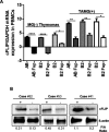
Figure 1
Figure 1
cFLIP mRNA and protein expression analysis by Q-RT-PCR and western blot. (A) mRNA expression in whole tissue extracts of AB thymomas (10 TAMG(+) and 16 (MG(−)), B2 thymomas (16 TAMG(+) and (13 MG(−)) and B3 thymomas (seven TAMG+ and 10 MG(−)) (+)); ****P < 10−4, ***P = 0.0002). (B) Protein expression in three TAMG(+) and three MG(−) AB thymomas. F/A denotes the ratio of the intensity values of cFLIP band and the respective actin (loading control) band as measured by densitometry using ImageJ program. cFLIP, cellular FLICE-like inhibitory protein; TAMG, thymoma-associated myasthenia gravis.
Figure 2
Figure 2
cFLIP mRNA and protein expression in thymocytes and PBMCs from TAMG(+) and MG(−) type AB and B2 thymomas. (A) Expression of mRNA in thymocytes from three TAMG(+) and three MG(−) type AB thymomas and five TAMG(+) and four MG(−) type B2 thymomas. (B) Expression of mRNA in PBMCs from patients with eight TAMG(+) and nine MG(−) type AB thymomas and five TAMG(+) and five MG(−) type B2 thymomas. (C and D) Protein expression in thymocytes and PBMCs from patients with type AB thymomas (three TAMG(+) and three MG(−) thymomas). F/A denotes the ratio of the intensity values of cFlip band and the respective actin (loading control) band as measured by densitometry using ImageJ program. cFLIP, cellular FLICE-like inhibitory protein; PBMCs, peripheral blood mononuclear cells; TAMG, thymoma-associated myasthenia gravis. *p = 0.020, 0.045, **p = 0.0077, 0.0071.
Figure 3
Figure 3
Intrapersonal comparison of cFLIP mRNA expression in thymoma (T) and the adjacent, non-neoplastic residual thymus (RT): six pairs each of TAMG(+) and MG(−) type AB thymomas and the respective RT from the same patients studied by qRT-PCR (**P = 0.0018, ***P = 0.0001 and ****P < 0.0001). cFLIP, cellular FLICE-like inhibitory protein; TAMG, thymoma-associated myasthenia gravis.
Figure 4
Figure 4
Expression of cFLIP in PBMCs and blood T-cell subsets. (A) PBMCs of blood donors (20–80 year-old people) (n = 20 in each decade), of patients with EOMG (anti-AChR-positive MG patients [25–39] years old, n = 9), MG(−) thymomas (n = 6; five type AB, one type B2) and TAMG(+) thymomas (n = 6; three AB, and three B2 [****p < 0.0001]). (B–D) cFLIP mRNA expression in blood-derived CD4(+) and CD8 (+) T cells and B cells from blood donors and patients with EOMG, MG(−) thymomas, and TAMG(+) thymomas (***P = 0.0010, **P = 0.0019 and 0.0095) and *P = 0.0286. (E) cFLIP mRNA levels in different T-cell subsets, CD4+CD45A(+), and CD45A(−) versus CD8+CD45A(+), and CD45A(−) from three blood donors (51–78 years old), six AB thymomas (three MG(−) and three TAMG(+) [55–71 years old]). (***P = 0.0006, **P = 0.0029, *P = 0.018 or P = 0.025, DP, double-positive; NS, not significant). GAPDH was used as reference for cFLIP mRNA analysis by real-time PCR. cFLIP, cellular FLICE-like inhibitory protein; PBMCs, peripheral blood mononuclear cells; EOMG, early onset myasthenia gravis; TAMG, thymoma-associated myasthenia gravis.
Figure 5
Figure 5
(A) cFLIP mRNA expression analysis by real-time PCR in PBMCs from two patients with type AB (#10 and #25) and four type B2 thymomas (#39, #41, #52, and #53) before thymectomy and on follow-up (Fup). ****P < 10−4 and **P = 0.0015, and 0.0044, *P = 0.012. (B) Analysis of cFLIP protein expression by western blot in two MG(−) type B2 thymomas (cases #52 and #53) and one TAMG(+) type B2 thymoma (#41). F/A denotes the ratio of the intensity values of the cFLIP band and the respective actin (loading control) band as measured by densitometry using ImageJ program. Fup was 2 months in case #41, 6 months in cases #10, #25, #52, and (#53); and 12 months in case #39. cFLIP, cellular FLICE-like inhibitory protein; PBMCs, peripheral blood mononuclear cells; TAMG, thymoma-associated myasthenia gravis.
Figure 6
Figure 6
Survival analysis of PBMCs from blood donors (n = 16, age range 20–80 years) and thymoma patients (n = 10; four MG(−) [two AB and two B2 subtype] and six TAMG(+) [three AB, two B2, and one B3 subtypes]) by MTT assay after treatment with FASLG (5 and 10 ng/mL for 24 h). There was a significant difference between thymoma patients and blood donors but not between TAMG(+) and MG(−) thymoma patients. Shown is one representative experiment (of two independent investigations; each assay was run in triplicate). PBMCs, peripheral blood mononuclear cells; TAMG, thymoma-associated myasthenia gravis; FASLG, Fas Ligand.
Figure 7
Figure 7
Impact of the NF-κB inhibitor EF24 on cFLIP expression of PBMCs from thymoma patients and blood donors. (A) QRT-PCR analysis of cFLIP expression in PBMCs from blood donors (n = 4) and five thymoma patients (two type AB, two B2, and one B3 thymoma). Left columns represent cFLIP mRNA levels in untreated PBMCs (cont); middle colums represent cFLIP levels in PBMCs treated with FASLG (10 ng/mL); right colums represent levels after pretreatment with EF24 for 24 h and subsequent FASLG treatment (10 ng/mL) for an additional 6 h. (B) cFLIP western blot analysis of whole PBMCs protein extracts (same samples and treatments as in [A]). 22, 35, 54, 61 50, 58, 66, 69, and 72 denote the ages of the patients (in years) from whom blood samples were obtained *P < 0.05, **P < 0.01, ***P < 0.001, and ****P < 10−4. cFLIP, cellular FLICE-like inhibitory protein; PBMCs, peripheral blood mononuclear cells; FASLG, Fas Ligand.
Figure 8
Figure 8
Impact of the NF-κB inhibitor EF24 on survival of PBMCs from thymoma patients and blood donors. (A) Cell survival analysis using the MTT assay of PBMCs from nine thymoma patients (four with AB, two with B2, and three B3 subtypes) treated either with EF24 alone or with EF24 followed by FASLG). (B) Apoptosis measurement by AnnexinV/APC/PI flow cytometry (FACS, Fluorescence-Activated Cell Sorting Guawa Millipore) in PBMCs of four blood donors and four thymoma patients. PBMCs were treated either with FASLG (10 ng/mL) or with EF24 for 24 h followed by FASLG (10 ng/mL) for an additional 6 h. 25, 39, 54, 61 50, 58, 66, and 72 denote the ages of the patients (in years) from whom blood sample were obtained. PBMCs, peripheral blood mononuclear cells; FASLG, Fas Ligand.

Similar articles

Cited by

References

    1. Müller-Hermelink HK, Engel PJ, Harris NL. Tumours of the thymus. In: Travis WD, Brambilla E, Müller-Hermelink HK, Harris CC, editors. Tumours of the lung, thymus, and heart pathology and genetics. Lyon: IARC Press; 2004. pp. 145–151. . In:, eds., et al..
    1. Marchevsky AM, Gupta R, McKenna RJ, et al. Evidence-based pathology and the pathologic evaluation of thymomas: the World Health Organization classification can be simplified into only 3 categories other than thymic carcinoma. Cancer. 2008;112:2780–2788. - PubMed
    1. Gupta R, Marchevsky AM, McKenna RJ, et al. Evidence-based pathology and the pathologic evaluation of thymomas: transcapsular invasion is not a significant prognostic feature. Arch Pathol Lab Med. 2008;132:926–930. - PubMed
    1. Huang B, Belharazem D, Li L, et al. Anti-apoptotic signature in thymic squamous cell carcinomas – functional relevance of anti-apoptotic BIRC3 expression in the thymic carcinoma cell line 1889c. Front Oncol. 2013;3:316. - PMC - PubMed
    1. Marx A, Pfister F, Schalke B, et al. The different roles of the thymus in the pathogenesis of the various myasthenia gravis subtypes. Autoimmun Rev. 2013;12:875–884. - PubMed

LinkOut - more resources