

Contributions of bilateral white matter to chronic aphasia symptoms as assessed by diffusion tensor MRI
- PMID: 26401977
- PMCID: PMC4669306
- DOI: 10.1016/j.bandl.2015.09.001
Contributions of bilateral white matter to chronic aphasia symptoms as assessed by diffusion tensor MRI
Abstract
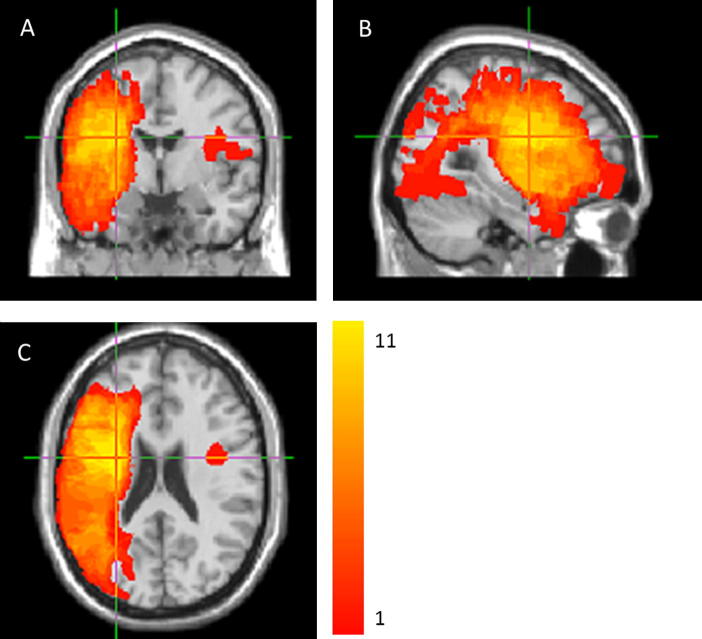
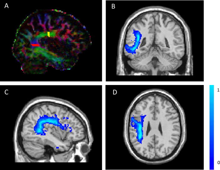
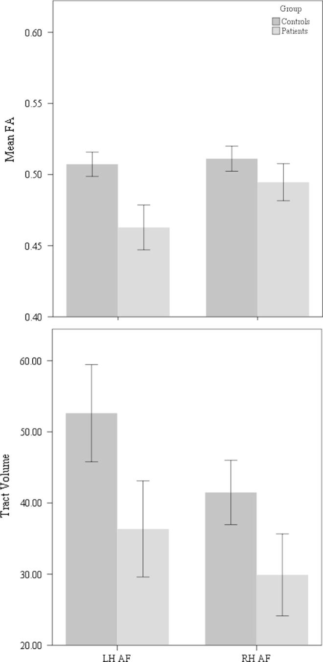
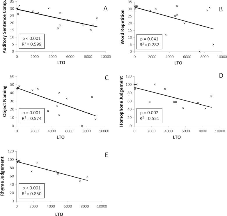
Language reorganisation following stroke has been studied widely. However, while studies of brain activation and grey matter examined both hemispheres, studies of white matter changes have mostly focused on the left hemisphere. Here we examined the relationship between bilateral hemispheric white matter and aphasia symptoms. 15 chronic stroke patients with aphasia and 18 healthy adults were studied using Diffusion Weighted Imaging data. By applying histogram analysis, Tract-Based Spatial Statistics, tractography and lesion-tract overlap methods, it was found that damage to the left hemisphere in general, and to the arcuate fasciculus in particular, correlated with impairments on word repetition, object naming, sentence comprehension and homophone and rhyme judgement. However, no such relationship was found in the right hemisphere. It is suggested that while some language function in aphasia can be explained by damage to the left arcuate fasciculus, it cannot be explained by looking at the contra-lesional tract.
Keywords: Arcuate fasciculus; Diffusion Tensor Imaging; Language; Stroke.
Copyright © 2015 The Authors. Published by Elsevier Inc. All rights reserved.
Figures
References
-
- Abel S., Weiller C., Huber W., Willmes K., Specht K. Therapy-induced brain reorganization patterns in aphasia. Brain. 2015;138:1097–1112. < http://brain.oxfordjournals.org/content/early/2015/02/14/brain.awv022#se...>. - PubMed
-
- Allendorfer J.B., Storrs J.M., Szaflarski J.P. Changes in white matter integrity follow excitatory rTMS treatment of post-stroke aphasia. Restorative Neurology and Neuroscience. 2012;30:103–113. < http://www.pubmedcentral.nih.gov/articlerender.fcgi?artid=3316910&tool=p...>. - PMC - PubMed
-
- Anglade C., Thiel A., Ansaldo A.I. The complementary role of the cerebral hemispheres in recovery from aphasia after stroke: A critical review of literature. Brain Injury. 2014;28:138–145. < http://informahealthcare.com/doi/full/10.3109/02699052.2013.859734>. - DOI - PubMed
-
- Benson D.F., Ardila A. Oxford University Press; Oxford: 1996. Aphasia: A clinical perspective.
-
- Berthier M.L., Lambon Ralph M.A., Pujol J., Green C. Arcuate fasciculus variability and repetition: The left sometimes can be right. Cortex. 2012;48:133–143. < http://www.sciencedirect.com/science/article/pii/S0010945211002024>. - PubMed
Publication types
MeSH terms
Grants and funding
LinkOut - more resources
Full Text Sources
Other Literature Sources
Medical
