Review
. 2015;48(2):319-53.
doi: 10.3233/JAD-142853.

Herpes Simplex Virus Type 1 and Other Pathogens are Key Causative Factors in Sporadic Alzheimer's Disease

Affiliations
Free PMC article
Review

Herpes Simplex Virus Type 1 and Other Pathogens are Key Causative Factors in Sporadic Alzheimer's Disease

Steven A Harris et al. J Alzheimers Dis. 2015.
Free PMC article

Abstract

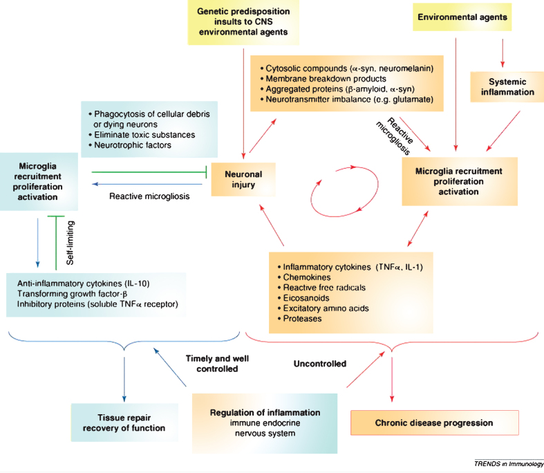
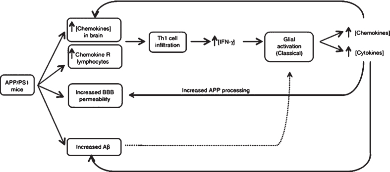
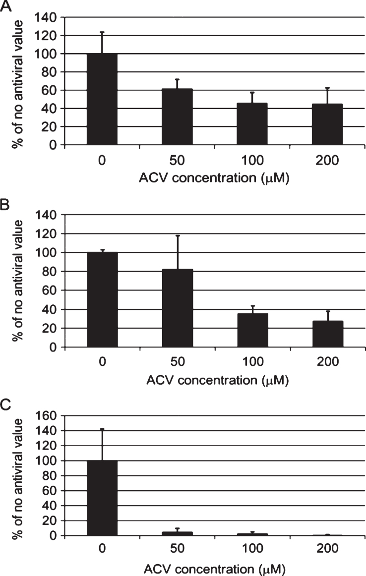
This review focuses on research in epidemiology, neuropathology, molecular biology, and genetics regarding the hypothesis that pathogens interact with susceptibility genes and are causative in sporadic Alzheimer's disease (AD). Sporadic AD is a complex multifactorial neurodegenerative disease with evidence indicating coexisting multi-pathogen and inflammatory etiologies. There are significant associations between AD and various pathogens, including Herpes simplex virus type 1 (HSV-1), Cytomegalovirus, and other Herpesviridae, Chlamydophila pneumoniae, spirochetes, Helicobacter pylori, and various periodontal pathogens. These pathogens are able to evade destruction by the host immune system, leading to persistent infection. Bacterial and viral DNA and RNA and bacterial ligands increase the expression of pro-inflammatory molecules and activate the innate and adaptive immune systems. Evidence demonstrates that pathogens directly and indirectly induce AD pathology, including amyloid-β (Aβ) accumulation, phosphorylation of tau protein, neuronal injury, and apoptosis. Chronic brain infection with HSV-1, Chlamydophila pneumoniae, and spirochetes results in complex processes that interact to cause a vicious cycle of uncontrolled neuroinflammation and neurodegeneration. Infections such as Cytomegalovirus, Helicobacter pylori, and periodontal pathogens induce production of systemic pro-inflammatory cytokines that may cross the blood-brain barrier to promote neurodegeneration. Pathogen-induced inflammation and central nervous system accumulation of Aβ damages the blood-brain barrier, which contributes to the pathophysiology of AD. Apolipoprotein E4 (ApoE4) enhances brain infiltration by pathogens including HSV-1 and Chlamydophila pneumoniae. ApoE4 is also associated with an increased pro-inflammatory response by the immune system. Potential antimicrobial treatments for AD are discussed, including the rationale for antiviral and antibiotic clinical trials.

Keywords: Alzheimer’s disease; ApoE4; Cytomegalovirus; Herpes simplex; amyloid; dementia; neurodegeneration; pathogen.


Figures

Fig.1
Fig.1
Co-localization of HSV-1 DNA and amyloid-β in AD plaques. A strong co-localization showing HSV-1 DNA (brown staining using PCR) and amyloid plaque (blue staining using immunohistochemistry) in a postmortem AD brain (G). Greater than 90% AD plaques contained viral DNA. Scale bar = 50μm. Figure from Wozniak, MA, Mee AP, Itzhaki RF (2009) Herpes simplex virus type 1 DNA is located within Alzheimer’s disease amyloid plaques. J Pathol 217, 131-138 [3]. Copyright 2008. Reprinted with permission from John Wiley and Sons, Inc. and Ruth Itzhaki.
Fig.2
Fig.2
Images demonstrating Chlamydophila pneumoniae in AD brain tissue by in situ hybridization. Figure (b) demonstrates C. pneumoniae from the hippocampus of an AD brain by in situ hybridization. Figures (d) and (e) show photographic enlargement of cells harboring C. pneumoniae inclusions identified in AD brain tissue. Arrows indicate the signal for C. pneumoniae. Image (b) was obtained using a x40 objective. Figure from Gérard HC, Dreses-Werringloer U, Wildt KS, Deka S, Oszust C, Balin BJ, Frey WH 2nd, Bordayo EZ, Whittum Hudson JA, Hudson AP (2006) Chlamydophila (Chlamydia) pneumoniae in the Alzheimer’s brain. FEMS Immunol Med Microbiol 48, 355-366 [7]. Copyright 2006. Reprinted with permission from John Wiley and Sons and permission from Brian Balin.
Fig.3
Fig.3
Association of spirochetes with Alzheimer’s disease. The frequency of spirochetes is significantly higher in the brains of Alzheimer’s disease patients compared to controls. Graph from Miklossy J (2011) Alzheimer’s disease - a neurospirochetosis. Analysis of the evidence following Koch’s and Hill’s criteria. J Neuroinflammation 8, 90 [126]. Copyright 2011. Reprinted with permission under the terms of the Creative Commons Attribution License, (http://creativecommons.org/licenses/by/2.0) and permission from Judith Miklossy.
Fig.4
Fig.4
Image of oral spirochetes in Alzheimer’s disease brain. The oral spirochete T. pectinovorum stained dark blue (arrows) in a section from the hippocampus from an 84-year-old woman with Alzheimer’s disease. The section was incubated with monoclonal antibodies to T. pectinovorum, and binding was disclosed using biotinylated anti-mouse antibodies and avidin-peroxidase. The photomicrograph was taken at1000X. Scale bar = 10μm. Figure from Riviere GR, Riviere KH, Smith KS (2002) Molecular and immunological evidence of oral Treponema in the human brain and their association with Alzheimer’s disease. Oral Microbiol Immunol 17, 113-118 [127]. Copyright 2002. Reprinted with permission from John Wiley and Sons, Inc. and George Riviere.
Fig.5
Fig.5
The vicious cycle of neurodegeneration. Neuroinflammation when controlled is reparative and self-limiting but when uncontrolled forms a vicious cycle and leads to chronic neurodegeneration. Figure from Gao HM, Hong JS (2008) Why neurodegenerative diseases are progressive: uncontrolled inflammation drives disease progression. Trends Immunol 29, 357-365 [1]. Copyright 2008. Reprinted with permission from Elsevier and John Hong.
Fig.6
Fig.6
The Lynch Hypothesis: T-cell lymphocytes infiltrate the brain and secrete IFN-γ which induces microglia activation and contributes to neurodegeneration in AD. Proposed sequence of events leading to amyloid pathology and microglial activation in AD. T lymphocytes cells activated peripherally cross the BBB and secrete IFN-γ and other cytokines, interact with microglia, and influence the neurodegenerative processes involved in AD. Figure from Lynch MA (2014) The impact of neuroimmune changes on development of amyloid pathology; relevance to Alzheimer’s disease. Immunology 141, 292-301 [167]. Copyright 2014. Reprinted with permission from John Wiley and Sons, Inc. andMarina Lynch.
Fig.7
Fig.7
Quantification of HSV-1 proteins (A), amyloid-β (B), and abnormal tau phosphorylation (C) in HSV-1-infected cells after acyclovir treatment. HSV-1 infected vero cell cultures treated with 0μM, 50μM, 100μM, or 200μM acyclovir (ACV). ACV significantly inhibited replication of HSV-1 as shown by a decrease in HSV-1 proteins (A). Aβ in cell cultures was reduced by 70% at a 200μM concentration of acyclovir (B). Abnormal tau phosphorylation was reduced nearly 100% at a 200μM concentration of acyclovir (C). p <  0.0001 compared to controls at all ACV concentrations for A and C and at 100μM and 200μM ACV concentrations for B. Graph from Wozniak MA et al. (2011) Antivirals reduce the formation of key Alzheimer’s disease molecules in cell cultures acutely infected with Herpes simplex virus type 1. PLoS One 6, e25152 [166]. Copyright 2011. Reprinted under the terms of the Creative Commons Attribution License, (http://creativecommons.org/licenses/by/2.0) and permission from Ruth Itzhaki.

References

    1. Gao HM, Hong JS. Why neurodegenerative diseases are progressive: Uncontrolled inflammation drives disease progression. Trends Immunol. 2008;29:357–365. - PMC - PubMed
    1. De-Paula VJ, Radanovic M, Diniz BS, Forlenza OV. Alzheimer’s disease. Subcell Biochem. 2012;65:329–352. - PubMed
    1. Wozniak MA, Mee AP, Itzhaki RF. Herpes simplex virus type 1 DNA is located within Alzheimer’s disease amyloid plaques. J Pathol. 2009;217:131–138. - PubMed
    1. Jamieson GA, Maitland NJ, Wilcock GK, Craske J, Itzhaki RF. Latent herpes simplex virus type 1 in normal and Alzheimer’s disease brains. J Med Virol. 1991;33:224–227. - PubMed
    1. Miklossy J. Emerging roles of pathogens in Alzheimer disease. Expert Rev Mol Med. 2011;13:e30. - PubMed