. 2015 Oct;21(10):1777-83.
doi: 10.3201/eid2110.150259.

Delayed Disease Progression in Cynomolgus Macaques Infected with Ebola Virus Makona Strain

Delayed Disease Progression in Cynomolgus Macaques Infected with Ebola Virus Makona Strain

Andrea Marzi et al. Emerg Infect Dis. 2015 Oct.

Abstract

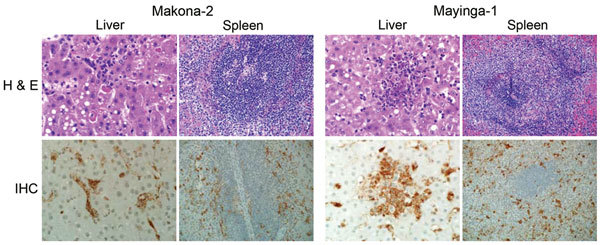
In late 2013, the largest documented outbreak of Ebola hemorrhagic fever started in Guinea and has since spread to neighboring countries, resulting in almost 27,000 cases and >11,000 deaths in humans. In March 2014, Ebola virus (EBOV) was identified as the causative agent. This study compares the pathogenesis of a new EBOV strain, Makona, which was isolated in Guinea in 2014 with the prototype strain from the 1976 EBOV outbreak in the former Zaire. Both strains cause lethal disease in cynomolgus macaques with similar pathologic changes and hallmark features of Ebola hemorrhagic fever. However, disease progression was delayed in EBOV-Makona-infected animals, suggesting decreased rather than increased virulence of this most recent EBOV strain.

Keywords: Ebola hemorrhagic fever; Ebola virus; Makona strain; Mayinga strain; West African Ebola virus isolate; cynomolgus macaques; nonhuman primates; pathogenesis; viruses.


Figures

Figure 1
Figure 1
Clinical parameters for 6 cynomolgus macaques infected with Ebola virus strains Makona or Mayinga. Parameters were measured for each animal on day of examination and time of euthanasia. A) Temperature profiles. B) Virus titer (viremia). TCID50, 50% tissue culture infectious dose. C) Daily clinical scorel. D) Survival curves showing significant difference in time to death between both groups (p = 0.0295, by Mantel-Cox test).
Figure 2
Figure 2
Blood and serum parameters for 6 cynomolgus macaques infected with Ebola virus strains Makona or Mayinga. Parameters were measured for each animal on day of examination and time of euthanasia. A) Leukocyte counts. WBC, white blood cell. B) Neutrophil (NE) counts. C) Platelet counts. D) Bilirubin levels. E) Aspartate aminotransferase (AST) levels. F) Akaline phosphatase (ALP) levels.
Figure 3
Figure 3
Serum cytokine and chemokine levels for 6 cynomolgus macaques infected with Ebola virus strains Makona or Mayinga. A) Tumor necrosis factor-α (TNF-α); B) Interleukin-6 (IL-6); C) Interferon-γ (IFN-γ); D) Soluble CD40 ligand (sCD40L); E) IL-10; and F) Monocyte chemotactic protein 1 (MCP-1). Kinetics were analyzed in serum samples of each animal collected on days of examination and time of euthanasia.
Figure 4
Figure 4
Virologic and pathologic results for 6 cynomolgus macaques infected with Ebola virus strains Makona or Mayinga. A) Viral infectivity titers were determined in key tissue samples collected at the time of euthanasia. TCID50, 50% tissue culture infectious dose; LN, lymph node. B) Pathology scores for liver and spleen. Scores were generated by using the scoring system described in the Materials and Methods.
Figure 5
Figure 5
Pathologic results for 6 cynomolgus macaques infected with Ebola virus strains Makona or Mayinga. Liver and spleen sections were stained with hematoxylin and eosin (H & E; top panels) and analyzed for necrosis, microthrombi, lymphocytosis, and inflammation. Sections were also stained with a polyclonal rabbit serum against Ebola virus viral protein 40 for detection of viral antigen (immunohistochemical [IHC] analysis; bottom panels). Sections from a representative animal in each group are shown. Original magnification levels: liver, ×40; spleen, ×20.

References

    1. Feldmann H, Geisbert TW. Ebola haemorrhagic fever. Lancet. 2011;377:849–62 and. 10.1016/S0140-6736(10)60667-8 - DOI - PMC - PubMed
    1. Baize S, Pannetier D, Oestereich L, Rieger T, Koivogui L, Magassouba N, et al. Emergence of Zaire Ebola virus disease in Guinea. N Engl J Med. 2014;371:1418–25 . 10.1056/NEJMoa1404505 - DOI - PubMed
    1. World Health Organization. Ebola situation report, May 13, 2015. Geneva: The Organization; 2015.
    1. Brining DL, Mattoon JS, Kercher L, LaCasse RA, Safronetz D, Feldmann H, et al. Thoracic radiography as a refinement methodology for the study of H1N1 influenza in cynomologus macaques (Macaca fascicularis). Comp Med. 2010;60:389–95 . - PMC - PubMed
    1. Reed LJ, Muench H. A simple method of estimating fifty percent endpoints. Am J Hyg. 1938;27:493–7.

Publication types