Observational Study
. 2015 Sep;94(38):e1547.
doi: 10.1097/MD.0000000000001547.

Desmoid-Type Fibromatosis of the Thorax: CT, MRI, and FDG PET Characteristics in a Large Series From a Tertiary Referral Center

Affiliations
Observational Study

Desmoid-Type Fibromatosis of the Thorax: CT, MRI, and FDG PET Characteristics in a Large Series From a Tertiary Referral Center

Hai Xu et al. Medicine (Baltimore). 2015 Sep.

Abstract

The purpose of this study was to describe the radiologic findings of computed tomography (CT), magnetic resonance (MR) imaging, and ¹⁸F-fluorodeoxy glucose positron emission tomography (FDG PET) in desmoid-type fibromatosis of the thorax. We retrospectively evaluated 47 consecutive patients with pathologically proven desmoid-type fibromatosis from January 2005 to March 2015. Patients underwent CT (n = 36) and/or MR (n = 32), and 13 patients also underwent FDG PET. Based on CT and MR, the sizes, locations, margins, contours, presence of surrounding fat, extra-compartment extension, bone involvement, and neurovascular involvement of the tumors were recorded. The attenuation, signal intensity, enhancement pattern, and presence of internal low signal band or signal void of the tumors were evaluated. Initial image findings were then compared between 2 groups of tumors: group 1 with recurrence or progression, and group 2 with no recurrence or stable without treatment. Median age at diagnosis of the tumors was 45 years, range 4 to 96, female-to-male ratio 1.8. Median tumor long diameter was 65 mm (range, 22-126 mm). The most common locations were chest wall (42.6%), followed by supraclavicular area, shoulder or axillary area, and mediastinum. The tumors had well-defined margins (83.0%), lobulated in contours (66.0%) surrounding fat (63.8%), extra-compartment extensions (42.6%), bone involvements (42.6%), and neurovascular involvements (27.7%). On CT, tumors had low attenuation (60.0%) with mild enhancement (median 24 HU, range 0-52). On MR, they showed iso-signal intensity (SI) (96.9%) on T1-weighted images (WI), and high SI (90.6%) on T2WI images, with strong (87.5%) and heterogeneous (96.9%) enhancement. Internal low signal bands (84.4%) and signal voids (68.8%) were noted. The median value of maxSUV was 3.1 (range, 2.0-7.3). In group 1 (n = 19, 40.4%), 13 patients suffered recurrence and 6 experienced progression. Group 2 (n = 28, 59.6%) consisted of 21 patients with no recurrence and 7 stable patients receiving no treatment. Partially ill-defined margins (OR, 0.167; 95% CI 0.029-0.943; P = 0.043) was the independent predictor for recurrence or progression of tumor. Knowledge of the radiological findings in desmoid-type fibromatosis on CT, MR, and FDG PET may help to improve diagnosis. Tumors with partially ill-defined margins have a tendency to recur or progress.


Conflict of interest statement

The authors have no funding and conflicts of interest to disclose.

Figures

FIGURE 1
FIGURE 1
Flow chart of patient selection.
FIGURE 2
FIGURE 2
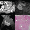
A 52-year-old man with desmoid-type fibromatosis in the chest wall without recurrence (Group 2). This was misdiagnosed as a neurogenic tumor based on preoperative CT but as fibromatosis based on preoperative MR. Axial pre- (A) and contrast-enhanced (B) CT axial images (5-mm reconstruction) show a circumscribed, well-defined mass () without enhancement in the right chest wall. Note the osteoblastic change of the rib (arrow). FDG PET reveals a hypometabolic mass, with a maximal standardized uptake value of 2.3 (not shown). (C) A mass shows abutting the rib () with no evidence of invasion of the cortex of bone and periosteum (arrows, ×40 magnification). CT = computed tomography, MR = magnetic resonance, FDG PET = 18F-fluorodeoxy glucose positron emission tomography.
FIGURE 3
FIGURE 3
A 47-year-old man presented with desmoid-type fibromatosis in the left supraclavicular area with recurrence (Group 1). It was misdiagnosed as a pancoast tumor due to lung cancer and a sarcoma of mesenchymal origin, based on preoperative CT and MR, respectively. (A) The contrast-enhanced axial (A) CT image (5-mm reconstruction) shows a huge, lobulated, partially ill-defined mass in the supraclavicular area (). Note the osteoblastic change of the first and second ribs (white arrows) and suspicion of involvement of the left subclavian artery (black arrow). Note the strong and heterogeneous enhancement with central areas of nonenhancing low signal bands (white arrows) and internal signal voids (black arrow) on enhanced T1-weighted axial (B) and sagittal (C) images. FDG PET revealed a hypermetabolic mass with a maximal standardized uptake value of 4.1 (not shown). (D) The mass was excised. Pathologically, the mass has infiltrated the brachial plexus, and tumor cells surround the nerve bundles (arrows, ×12.5 magnification). CT = computed tomography, MR = magnetic resonance, FDG PET = 18F-fluorodeoxy glucose positron emission tomography.
FIGURE 4
FIGURE 4
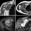
A 47-year-old woman with desmoid-type fibromatosis in the right axilla with progression (Group 1). This was misdiagnosed as a sarcoma on preoperative CT but as fibromatosis on preoperative MR. The tumor shows iso-signal intensity on coronal T1-weighted image (A) and high signal intensity on axial T2-weighted image (B). The margin is partially ill-defined (white arrows) on axial T2-weighted image (B). A rim of surrounding fat (white arrows) with high signal intensity is clearly seen on T1-weighted image (A). On enhanced T1-weighted axial (C) and coronal (D) images, the tumor shows strong and heterogeneous enhancement with a central area of nonenhancing low signal bands (black arrows). The maximal standardized uptake value on FDG PET was 2.1 (not shown). CT = computed tomography, MR = magnetic resonance, FDG PET = 18F-fluorodeoxy glucose positron emission tomography.
FIGURE 5
FIGURE 5
A 43-year-old man with desmoid-type fibromatosis in the mediastinum with recurrence (Group 1). This was misdiagnosed as a neurogenic tumor and as fibrosarcoma on preoperative CT and MR, respectively. (A) The contrast-enhanced axial CT image (5-mm reconstruction) shows a lobulated, well-defined mass () with mild homogeneous enhancement in the left posterior mediastinum. The difference in Hounsfield units between pre- and postenhancement is 7. Note the osteoblastic change of the rib (white arrow) and the neural foraminal widening (black arrow). (B) The tumor shows strong heterogeneous enhancement on an enhanced T1-weighted image. Note the internal nonenhancing low signal bands (white arrows) and internal signal voids (black arrows). (C) Grossly, an ill-defined irregularly-shaped firm mass is present. The cut surface of the mass glistens, and is whitish gray, coarsely trabeculated and partly myxoid, without hemorrhage and necrosis. (D) Tumor cells infiltrate the chest wall muscle (white arrows, ×40 magnification) and (E) also the marrow space of the rib (, ×100 magnification). CT = computed tomography, MR = magnetic resonance.

References

    1. Weiss SW, Goldblum JR, Flope AL. Enzinger and Weiss's Soft Tissue Tumors: Expert Consults. 6th ed.2014; Philadelphia: Saunders Elsevier, 288–293.
    1. Fletcher CD, Unni KK, Mertens F. Pathology and Genetics of Tumours of Soft Tissue and Bone. 2002; Lyon: IARC Press, 83.
    1. Hosalkar HS, Torbert JT, Fox EJ, et al. Musculoskeletal desmoid tumors. J Am Acad Orthop Surg 2008; 16:188–198. - PubMed
    1. Nuyttens JJ, Rust PF, Thomas CR, Jr, et al. Surgery versus radiation therapy for patients with aggressive fibromatosis or desmoid tumors: a comparative review of 22 articles. Cancer 2000; 88:1517–1523. - PubMed
    1. Janinis J, Patriki M, Vini L, et al. The pharmacological treatment of aggressive fibromatosis: a systematic review. Ann Oncol 2003; 14:181–190. - PubMed

Publication types

MeSH terms

Substances