

Circulating Tumor Cell Analyses in Patients With Esophageal Squamous Cell Carcinoma Using Epithelial Marker-Dependent and -Independent Approaches
- PMID: 26402816
- PMCID: PMC4635756
- DOI: 10.1097/MD.0000000000001565
Circulating Tumor Cell Analyses in Patients With Esophageal Squamous Cell Carcinoma Using Epithelial Marker-Dependent and -Independent Approaches
Abstract
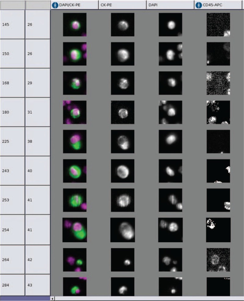
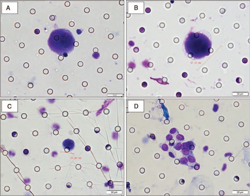
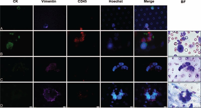
In several epithelial malignancies, detection of circulating tumor cells (CTCs) in the peripheral blood has diagnostic, prognostic, and therapeutic implications. However, the clinical relevance of CTCs in esophageal squamous cell carcinoma (ESCC) has not yet been ascertained. The study was conducted with the aim of determining the clinical significance of CTCs in patients with ESCC by using 2 CTC detection systems, one epithelial marker-dependent and the other epithelial marker-independent. Paired peripheral blood samples were prospectively obtained from 61 ESCC patients before treatment and were analyzed for CTCs isolated by the CellSearch system (CS) and the method of isolation by size of epithelial tumor (ISET). Blood samples from 22 healthy volunteers were used as controls. Out of 61 study subjects, CTCs were detected in 20 patients (32.8%) by the ISET method and in only 1 patient (1.6%) by the CS method. Circulating tumor microemboli (CTM) were observed in 3 of 61 (4.9%) patients using ISET, but were undetectable in any of the patient by CS method. No CTCs/CTM were detected by either method in control groups. By ISET method, the presence of CTCs appeared to correlate with the stage of ESCC and with the baseline median platelet levels. No correlation with any other relevant clinicopathological variables was observed. Our results clearly indicate the ability of both CS and ISET methods to detect CTCs in peripheral blood samples from ESCC patients. However, the CellSearch system appears to have a poorer sensitivity as compared with the ISET method. Further studies are essential for assessing the role of such technologies in ESCC.
Conflict of interest statement
The authors have no conflicts of interest to disclose.
Figures
References
-
- Ferlay J, Soerjomataram I, Dikshit R, et al. Cancer incidence and mortality worldwide: sources, methods and major patterns in GLOBOCAN 2012. Int J Cancer 2015; 136:E359–E386. - PubMed
-
- Kamangar F, Dores GM, Anderson WF. Patterns of cancer incidence, mortality, and prevalence across five continents: defining priorities to reduce cancer disparities in different geographic regions of the world. J Clin Oncol 2006; 24:2137–2150. - PubMed
-
- Pennathur A, Gibson MK, Jobe BA, et al. Oesophageal carcinoma. Lancet 2013; 381:400–412. - PubMed
-
- Paterlini-Brechot P, Benali NL. Circulating tumor cells (CTC) detection: clinical impact and future directions. Cancer Lett 2007; 253:180–204. - PubMed
-
- Allard WJ, Matera J, Miller MC, et al. Tumor cells circulate in the peripheral blood of all major carcinomas but not in healthy subjects or patients with nonmalignant diseases. Clin Cancer Res 2004; 10:6897–6904. - PubMed
Publication types
MeSH terms
Substances
LinkOut - more resources
Full Text Sources
Medical
