

Close reduction and percutaneous pinning in displaced supracondylar humerus fractures in children
- PMID: 26403444
- PMCID: PMC3872817
- DOI: 10.1016/j.jcot.2012.09.004
Close reduction and percutaneous pinning in displaced supracondylar humerus fractures in children
Abstract
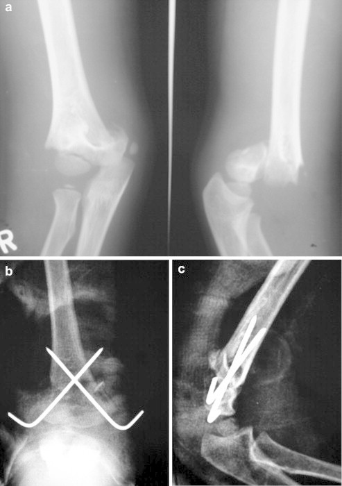
Background: Displaced supracondylar fractures of the humerus in children are common pediatric injuries treated by orthopedic surgeons. They also have a high rate of complications if not reduced and stabilized in optimal position which may lead to serious neurovascular injuries and residual deformity. Amongst the various methods used for treating these fractures, closed reduction and percutaneous pinning has shown improved results.
Method: Between March 2005 and April 2010, 277 cases of supracondylar humeral fractures (Gartland grade II and III) with less then 1 week old were included in this study. They were treated with closed reduction and percutaneous pinning with crossed Kirschner wires under image intensifier control. Clinical outcome were assessed according to Flynn's criteria.
Results: The mean age at the time of operation was 6 years (range 2-10 years) and the average duration of follow-up was 4.6 years (range 2.1-7.2 years). The Flynn's criteria were excellent in 202, good in 68, fair in 5 and only 2 with poor results.
Conclusion: Closed reduction and percutaneous pinning is a sound and effective treatment for displaced supracondylar fractures.
Keywords: Closed reduction; Displaced supracondylar fractures; Percutaneous pinning.
Figures


Similar articles
-
Clinical Results of Closed Reduction and Percutaneous Pinning for Gartland Type II Flexion-Type Supracondylar Humeral Fractures in Children: Report of Three Cases.J Nippon Med Sch. 2023;90(3):294-300. doi: 10.1272/jnms.JNMS.2023_90-402. J Nippon Med Sch. 2023. PMID: 37380478
-
Displaced supracondylar humeral fractures in children: Comparison of three treatment approaches.Srp Arh Celok Lek. 2016 Jan-Feb;144(1-2):46-51. doi: 10.2298/sarh1602046d. Srp Arh Celok Lek. 2016. PMID: 27276857
-
Closed reduction and percutaneous pinning of displaced supracondylar fractures of humerus in children with delayed presentation.Chin J Traumatol. 2011 Feb 1;14(1):14-9. Chin J Traumatol. 2011. PMID: 21276362
-
Management of pediatric distal humerus metaphyseal-diaphyseal junction fracture: A systematic review and meta-analysis.J Child Orthop. 2024 Jun 22;18(4):421-431. doi: 10.1177/18632521241262169. eCollection 2024 Aug. J Child Orthop. 2024. PMID: 39100985 Free PMC article. Review.
-
[Supracondylar fractures of the humerus in children].Tidsskr Nor Laegeforen. 2011 Feb 18;131(4):349-52. doi: 10.4045/tidsskr.10.0428. Tidsskr Nor Laegeforen. 2011. PMID: 21339783 Review. Norwegian.
Cited by
-
Kirschner Wire Prying and Leverage Technique: a new closed reduction method in treatment of pediatric "Irreducible Supracondylar Humerus Fractures".J Orthop Surg Res. 2024 Feb 2;19(1):113. doi: 10.1186/s13018-024-04592-4. J Orthop Surg Res. 2024. PMID: 38308347 Free PMC article.
-
Treatment of grossly dislocated supracondylar humerus fractures after failed closed reduction: a retrospective analysis of different surgical approaches.Arch Orthop Trauma Surg. 2022 Aug;142(8):1933-1940. doi: 10.1007/s00402-021-03937-6. Epub 2021 May 13. Arch Orthop Trauma Surg. 2022. PMID: 33983529
-
Neurovascular complications after supracondylar humerus fractures in children.Trauma Case Rep. 2017 Jan 6;8:16-19. doi: 10.1016/j.tcr.2017.01.013. eCollection 2017 Apr. Trauma Case Rep. 2017. PMID: 29644308 Free PMC article.
-
Leverage application on Gartland type IV supracondylar humeral fracture in children.Int Orthop. 2016 Nov;40(11):2417-2422. doi: 10.1007/s00264-016-3206-3. Epub 2016 May 26. Int Orthop. 2016. PMID: 27230232
-
Supracondylar Humerus Fractures: Classification Based Treatment Algorithms.Indian J Orthop. 2020 Oct 22;55(1):68-80. doi: 10.1007/s43465-020-00285-2. eCollection 2021 Feb. Indian J Orthop. 2020. PMID: 33569100 Free PMC article. Review.
References
-
- Mostafavi H.R., Spero C. Crossed pin fixation of displaced supracondylar humerus fractures in children. Clin Orthop Relat Res. 2000;376:56–61. - PubMed
-
- D'Ambrosia R.D. Supracondylar fractures of humerus – prevention of cubitus varus. J Bone Jt Surg Am. 1972;54:60–66. - PubMed
-
- Aronson D.D., Prager B.I. Supracondylar fractures of the humerus in children. A modified technique for closed pinning. Clin Orthop Relat Res. 1987;219:174–184. - PubMed
-
- Gartland J.J. Management of supracondylar fractures in children. Surg Gynecol Obstet. 1959;109:145–154. - PubMed
-
- Dunlop J. Transcondylar fracture of the humerus in children. J Bone Jt Surg Am. 1939;21:59–73.
LinkOut - more resources
Full Text Sources
Miscellaneous
