. 2015 Sep 24:15:185.
doi: 10.1186/s12866-015-0516-3.

Breakdown of albumin and haemalbumin by the cysteine protease interpain A, an albuminase of Prevotella intermedia

Affiliations

Breakdown of albumin and haemalbumin by the cysteine protease interpain A, an albuminase of Prevotella intermedia

Dominic P Byrne et al. BMC Microbiol. .

Abstract

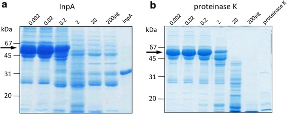
Background: Prevotella intermedia is a Gram-negative black-pigmenting oral anaerobe associated with periodontitis in humans, and has a haem requirement for growth, survival and virulence. It produces an iron porphyrin-containing pigment comprising monomeric iron (III) protoporphyrin IX (Fe(III)PPIX.OH; haematin). The bacterium expresses a 90-kDa cysteine protease termed interpain A (InpA) which both oxidizes and subsequently degrades haemoglobin, releasing haem. However, it is not known whether the enzyme may play a role in degrading other haem-carrying plasma proteins present in the gingival sulcus or periodontal pocket from which to derive haem. This study evaluated the ability of InpA to degrade apo- and haem-complexed albumin.

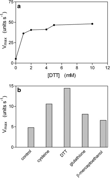
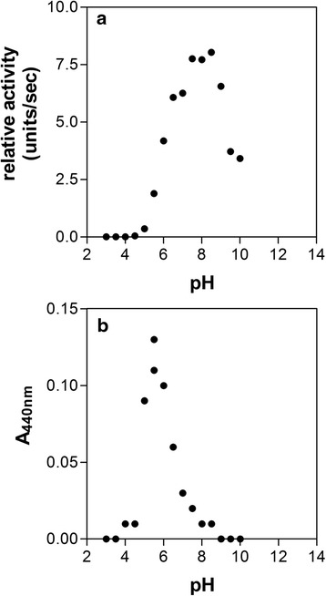
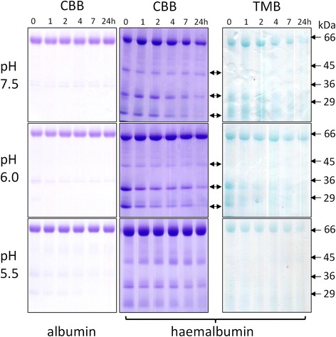
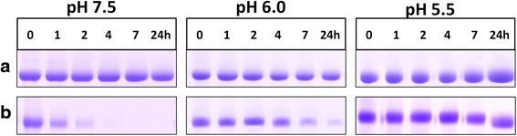
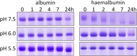
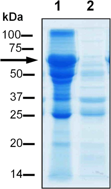
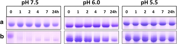
Results: Albumin breakdown was examined over a range of pH and in the presence of reducing agent; conditions which prevail in sub- and supra-gingival plaque. InpA digested haemalbumin more efficiently than apoalbumin, especially under reducing conditions at pH 7.5. Under these conditions InpA was able to substantially degrade the albumin component of whole human plasma.

Conclusions: The data point to InpA as an efficient "albuminase" with the ability to degrade the minor fraction of haem-bound albumin in plasma. InpA may thus contribute significantly to haem acquisition by P. intermedia under conditions of low redox potential and higher pH in the inflamed gingival crevice and diseased periodontal pocket where haem availability is tightly controlled by the host.


Figures

Fig. 1
Fig. 1
Effect of dithiothreitol (a) and other thiol reducing agents (b) on the enzyme activity of InpA. The enzyme activity was assayed in triplicate using Z-Arg-Arg-AMC at 37 °C in 0.1M Tris–HCl, pH 7.5 and the concentrations of the reducing agents in Fig. 1b were 3 mM. Due to the high reproducibility of the replicates, no SD is apparent. Control, activity in the absence of reducing agent. InpA concentrations in (a) and (b) were 12.5 μg ml−1. See text for details
Fig. 2
Fig. 2
Effect of pH on InpA activity towards Z-Arg-Arg-AMC (a) and azoalbumin (b). Protease activity was assayed at 37 °C in PBS, pH 7.4, containing 3 mM DTT. InpA concentration was 50 μg ml−1. Azoalbumin concentration was 3 % (w/v). Enzyme activity is depicted as absorbance of azo dye (A440) released from the protein. See text for details
Fig. 3
Fig. 3
Albumin and haemalbumin breakdown by InpA at different pH under non-reducing conditions. Albumin and haemalbumin (1:1 haem:protein ratio) (16 μM) were incubated with InpA (100 μg ml−1) at 37 °C and aliquots of the incubations were sampled periodically at 1, 2, 4, 7 and 24 h and InpA activity inhibited by E-64 (0.5 mM) and subjected to SDS-PAGE on 10 % non-reducing gels. Samples were solubilised at 37 °C for 1 h in non-reducing application buffer. The haemalbumin gels were stained with TMB to reveal haem-associated peroxidise activity and counterstained with CBB for protein, whilst albumin gels were stained only with CBB. Protein loading was ≈ 20 μg per lane. Arrows denote digestion fragments carrying bound haem
Fig. 4
Fig. 4
Chemical reduction increases the susceptibility of albumin to InpA. Albumin (16 μM) was incubated with InpA (100 μg ml−1) at different pH at 37 °C under non-reducing (a panels) and reducing conditions (3 mM DTT) (b panels). Gel lanes had a nominal loading of 20 μg albumin, and are stained with CBB. Samples were prepared and gels run as described for Fig. 3
Fig. 5
Fig. 5
SDS-PAGE showing the stability of albumin and haemalbumin under reducing conditions at different pH. Albumin and haemalbumin (16 μM) were reduced with 3 mM DTT and incubated at 37 °C. SDS-PAGE conditions were the same as those in Fig. 3. The gels were stained with CBB
Fig. 6
Fig. 6
SDS-PAGE analysis of the breakdown of whole human plasma by InpA. Plasma (1 μl, ~60μg protein), was incubated overnight at 37 °C in PBS, pH 7.4, containing 3 mM DTT in absence (lane 1) or presence (lane 2) of active InpA (100 μg ml−1) in a final volume of 20 μl. ~ 50 μg total protein was loaded per lane. Arrow denotes albumin band. Samples were solubilised for 5 min at 100 °C in reducing application buffer, and gels were stained with CBB
Fig. 7
Fig. 7
Comparative SDS-PAGE analysis of the digestion of whole human plasma by InpA and proteinase K. Whole human plasma (60 μg total protein) was incubated overnight with InpA and proteinase K (Sigma-Aldrich; product number P2308) at the indicated concentrations (μg ml−1) at 37 °C in PBS, pH 7.4 containing 3 mM DTT. Last lane of each gel was loaded with 10 μg of either InpA or proteinase K only. Samples were prepared and gels stained as in Fig. 6
Fig. 8
Fig. 8
Intrinsic fluorescence spectra of bovine albumin in the presence of DTT. Albumin (16 μM) was treated with increasing concentrations of DTT in 0.14 M NaCl, 0.1 M Tris–HCl buffer, pH 7.5, at 37 °C, and the fluorescence emission monitored after excitation at 295 nm
Fig. 9
Fig. 9
Effect of reducing conditions on breakdown of haemalbumin by InpA at different pH. Haemalbumin (16 μM) with a 1:1 haem to protein ratio was incubated at 37 °C with InpA (100 μg ml−1) in the absence (a panels) and presence of 3 mM DTT (b panels). Samples and gels were run under non-reducing conditions on 10 % gels and stained for protein with CBB. Each gel lane had a nominal loading of 20 μg albumin
Fig. 10
Fig. 10
Primary sequence showing all the cleavage sites inflicted during incubation of bovine serum albumin with InpA. Albumin (16 μM) was incubated at 37 °C with InpA (100 μg ml−1) in 10 mM Tris buffer, pH 7.5, in the presence or absence of 2mM DTT, and the incubation mixture sampled over time subjected to MALDI-TOF mass spectrometry. InpA scission sites exclusive to reducing (formula image) , non-reducing (formula image), and to both incubation conditions (formula image) are indicated. See text for details

References

    1. Haffajee AD, Socransky SS. Microbial etiological agents of destructive periodontal diseases. Periodontol 2000. 1994;5:78–111. doi: 10.1111/j.1600-0757.1994.tb00020.x. - DOI - PubMed
    1. Smalley JW, Silver J, Birss AJ, Withnall R, Titler PJ. The haem pigment of the oral anaerobes Prevotella nigrescens and Prevotella intermedia is composed of iron (III) protoporphyrin IX in the monomeric form. Microbiology. 2003;149:1711–1718. doi: 10.1099/mic.0.26258-0. - DOI - PubMed
    1. Smalley JW, Birss AJ, Silver J. The periodontal pathogen Porphyromonas gingivalis harnesses the chemistry of the μ-oxo bishaem of iron protoporphrin IX to protect against hydrogen peroxide. FEMS Microbiol Lett. 2000;183:159–164. - PubMed
    1. Beaven GH, Chen SH, D’Albis A, Gratzer WD. A spectroscopic study of the haemin-human-serum-albumin system. Eur J Biochem. 1974;72:539–546. doi: 10.1111/j.1432-1033.1974.tb03295.x. - DOI - PubMed
    1. Hrkal Z, Vodrážka Z, Kalousek I. Transfer of haem from ferrihemoglobin and ferrihemoglobin isolated chains to hemopexin. Eur J Biochem. 1974;43:73–78. doi: 10.1111/j.1432-1033.1974.tb03386.x. - DOI - PubMed

Publication types

LinkOut - more resources