Multivalent Forms of the Notch Ligand DLL-1 Enhance Antitumor T-cell Immunity in Lung Cancer and Improve Efficacy of EGFR-Targeted Therapy
- PMID: 26404003
- PMCID: PMC4651735
- DOI: 10.1158/0008-5472.CAN-14-1154
Multivalent Forms of the Notch Ligand DLL-1 Enhance Antitumor T-cell Immunity in Lung Cancer and Improve Efficacy of EGFR-Targeted Therapy
Abstract
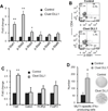
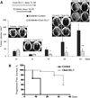
Activation of Notch signaling in hematopoietic cells by tumors contributes to immune escape. T-cell defects in tumors can be reversed by treating tumor-bearing mice with multivalent forms of the Notch receptor ligand DLL-1, but the immunologic correlates of this effect have not been elucidated. Here, we report mechanistic insights along with the efficacy of combinational treatments of multivalent DLL-1 with oncoprotein targeting drugs in preclinical mouse models of lung cancer. Systemic DLL-1 administration increased T-cell infiltration into tumors and elevated numbers of CD44(+)CD62L(+)CD8(+) memory T cells while decreasing the number of regulatory T cells and limiting tumor vascularization. This treatment was associated with upregulation of Notch and its ligands in tumor-infiltrating T cells enhanced expression of T-bet and phosphorylation of Stat1/2. Adoptive transfer of T cells from DLL1-treated tumor-bearing immunocompetent hosts into tumor-bearing SCID-NOD immunocompromised mice attenuated tumor growth and extended tumor-free survival in the recipients. When combined with the EGFR-targeted drug erlotinib, DLL-1 significantly improved progression-free survival by inducing robust tumor-specific T-cell immunity. In tissue culture, DLL1 induced proliferation of human peripheral T cells, but lacked proliferative or clonogenic effects on lung cancer cells. Our findings offer preclinical mechanistic support for the development of multivalent DLL1 to stimulate antitumor immunity.
©2015 American Association for Cancer Research.
Conflict of interest statement
Figures


Similar articles
-
Determinant roles of dendritic cell-expressed Notch Delta-like and Jagged ligands on anti-tumor T cell immunity.J Immunother Cancer. 2019 Apr 2;7(1):95. doi: 10.1186/s40425-019-0566-4. J Immunother Cancer. 2019. PMID: 30940183 Free PMC article.
-
Long-term engraftment and expansion of tumor-derived memory T cells following the implantation of non-disrupted pieces of human lung tumor into NOD-scid IL2Rgamma(null) mice.J Immunol. 2008 May 15;180(10):7009-18. doi: 10.4049/jimmunol.180.10.7009. J Immunol. 2008. PMID: 18453623
-
Resuscitating cancer immunosurveillance: selective stimulation of DLL1-Notch signaling in T cells rescues T-cell function and inhibits tumor growth.Cancer Res. 2011 Oct 1;71(19):6122-31. doi: 10.1158/0008-5472.CAN-10-4366. Epub 2011 Aug 8. Cancer Res. 2011. PMID: 21825014 Free PMC article.
-
Antitumor cytotoxic T-lymphocyte response in human lung carcinoma: identification of a tumor-associated antigen.Immunol Rev. 2002 Oct;188:114-21. doi: 10.1034/j.1600-065x.2002.18810.x. Immunol Rev. 2002. PMID: 12445285 Review.
-
Specific Targeting of Notch Ligand-Receptor Interactions to Modulate Immune Responses: A Review of Clinical and Preclinical Findings.Front Immunol. 2020 Aug 14;11:1958. doi: 10.3389/fimmu.2020.01958. eCollection 2020. Front Immunol. 2020. PMID: 32922403 Free PMC article. Review.
Cited by
-
Identification of important invasion and proliferation related genes in adrenocortical carcinoma.Med Oncol. 2019 Jul 18;36(9):73. doi: 10.1007/s12032-019-1296-7. Med Oncol. 2019. PMID: 31321566
-
Host-microbe computational proteomic landscape in oral cancer revealed key functional and metabolic pathways between Fusobacterium nucleatum and cancer progression.Int J Oral Sci. 2025 Jan 2;17(1):1. doi: 10.1038/s41368-024-00326-8. Int J Oral Sci. 2025. PMID: 39743544 Free PMC article.
-
Nanoparticles for Manipulation of the Developmental Wnt, Hedgehog, and Notch Signaling Pathways in Cancer.Ann Biomed Eng. 2020 Jul;48(7):1864-1884. doi: 10.1007/s10439-019-02399-7. Epub 2019 Nov 4. Ann Biomed Eng. 2020. PMID: 31686312 Free PMC article. Review.
-
Notching tumor: Signaling through Notch receptors improves antitumor T cell immunity.Oncoimmunology. 2016 Jan 11;5(5):e1122864. doi: 10.1080/2162402X.2015.1122864. eCollection 2016 May. Oncoimmunology. 2016. PMID: 27467934 Free PMC article.
-
Collective metastasis: coordinating the multicellular voyage.Clin Exp Metastasis. 2021 Aug;38(4):373-399. doi: 10.1007/s10585-021-10111-0. Epub 2021 Jul 12. Clin Exp Metastasis. 2021. PMID: 34254215 Free PMC article. Review.
References
-
- Fiuza UM, Arias AM. Cell and molecular biology of Notch. J Endocrinol. 2007;194:459–474. - PubMed
-
- Bettelli E, Carrier Y, Gao W, et al. Reciprocal developmental pathways for the generation of pathogenic effector TH17 and regulatory T cells. Nature. 2006;441:235–238. - PubMed
-
- Maekawa Y, Tsukumo S, Chiba S, et al. Delta1-Notch3 interactions bias the functional differentiation of activated CD4+ T cells. Immunity. 2003;19:549–559. - PubMed
Publication types
MeSH terms
Substances
Grants and funding
LinkOut - more resources
Full Text Sources
Other Literature Sources
Medical
Molecular Biology Databases
Research Materials
Miscellaneous

