. 2016 Jan;43(1):34-41.
doi: 10.1007/s00259-015-3188-1. Epub 2015 Sep 25.

(68)Ga-PSMA-11 PET/CT: a new technique with high potential for the radiotherapeutic management of prostate cancer patients

Affiliations

(68)Ga-PSMA-11 PET/CT: a new technique with high potential for the radiotherapeutic management of prostate cancer patients

Florian Sterzing et al. Eur J Nucl Med Mol Imaging. 2016 Jan.

Abstract

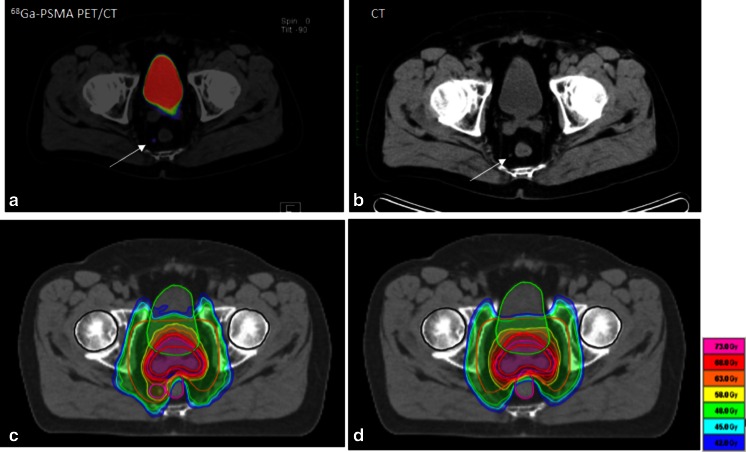
Purpose: Radiotherapy is the main therapeutic approach besides surgery of localized prostate cancer. It relies on risk stratification and exact staging. This report analyses the potential of [(68)Ga]Glu-urea-Lys(Ahx)-HBED-CC ((68)Ga-PSMA-11), a new positron emission tomography (PET) tracer targeting prostate-specific membrane antigen (PSMA) for prostate cancer staging and individualized radiotherapy planning.

Methods: A cohort of 57 patients with prostate cancer scanned with (68)Ga-PSMA-11 PET/CT for radiotherapy planning was retrospectively reviewed; 15 patients were at initial diagnosis and 42 patients at time of biochemical recurrence. Staging results of conventional imaging, including bone scintigraphy, CT or MRI, were compared with (68)Ga-PSMA ligand PET/CT results and the influence on radiotherapeutic management was quantified.

Results: (68)Ga-PSMA ligand PET/CT had a dramatic impact on radiotherapy application in the presented cohort. In 50.8 % of the cases therapy was changed.

Conclusion: The presented imaging technique of (68)Ga-PSMA PET/CT could be a key technology for individualized radiotherapy management in prostate cancer.

Keywords: 68Ga-PSMA ligand PET/CT; Individualized radiotherapy; Prostate cancer; Radiotherapy planning.


Figures

Fig. 1
Fig. 1
Overview of the impact of PSMA staging results on radiotherapy (RX). In light grey cases treatment was carried out as initially planned based on conventional staging information, and in dark grey cases radiotherapeutic management was changed in dose or target volume
Fig. 2
Fig. 2
Example of the impact of PSMA imaging on radiotherapeutic management at initial diagnosis of intermediate-risk prostate cancer. a PSMA PET/CT with tracer uptake in a pararectal lymph node (SUVmax 3.1; arrow) which was not clearly pathological in conventional CT (b). Accordingly the irradiation plan was changed with coverage of perirectal space and a simultaneous boost to the lymph node (c: IMRT in 34 fractions with 51 Gy to lymphatic pathways, 76.5 Gy to the prostate and 61.2 Gy to the pathological lymph node). d IMRT plan prior to PSMA PET information without sufficient coverage of the pathological lymph node
Fig. 3
Fig. 3
Prostate cancer recurrence in the intermediate-risk situation. a, b PSMA PET/CT with a pathological presacral lymph node that was unclear in conventional CT (c). The corresponding IMRT plan is shown in d with 34 fractions and 51 Gy to the lymphatic pathways (arrow) and a simultaneous integrated boost to the lymph node to 61.2 Gy. For comparison e displays the IMRT plan without PET information
Fig. 4
Fig. 4
Example of PSA relapse 3 years after prostatectomy and prostate bed irradiation for high-risk prostate cancer. Only the PSMA PET/CT (a) reveals the iliac lymph node metastases with an SUVmax of 9.7, whereas the conventional CT (b) could show nothing. The corresponding IMRT plan is shown in c with 26 fractions to the pelvic lymphatic pathways to 46.8 Gy and a simultaneous integrated boost to the lymph node to a dose of 57.2 Gy. After prostate bed irradiation 3 years before radiotherapy would not have been possible without the PET information

References

    1. Sooriakumaran P, Nyberg T, Akre O, Haendler L, Heus I, Olsson M, et al. Comparative effectiveness of radical prostatectomy and radiotherapy in prostate cancer: observational study of mortality outcomes. BMJ. 2014;348:g1502. doi: 10.1136/bmj.g1502. - DOI - PMC - PubMed
    1. Roethke M, Anastasiadis AG, Lichy M, Werner M, Wagner P, Kruck S, et al. MRI-guided prostate biopsy detects clinically significant cancer: analysis of a cohort of 100 patients after previous negative TRUS biopsy. World J Urol. 2012;30(2):213–218. doi: 10.1007/s00345-011-0675-2. - DOI - PubMed
    1. Kuru TH, Roethke MC, Seidenader J, Simpfendörfer T, Boxler S, Alammar K, et al. Critical evaluation of magnetic resonance imaging targeted, transrectal ultrasound guided transperineal fusion biopsy for detection of prostate cancer. J Urol. 2013;190(4):1380–1386. doi: 10.1016/j.juro.2013.04.043. - DOI - PubMed
    1. Pickett B, Vigneault E, Kurhanewicz J, Verhey L, Roach M. Static field intensity modulation to treat a dominant intra-prostatic lesion to 90 Gy compared to seven field 3-dimensional radiotherapy. Int J Radiat Oncol Biol Phys. 1999;44(4):921–929. doi: 10.1016/S0360-3016(98)00502-1. - DOI - PubMed
    1. Housri N, Ning H, Ondos J, Choyke P, Camphausen K, Citrin D, et al. Parameters favorable to intraprostatic radiation dose escalation in men with localized prostate cancer. Int J Radiat Oncol Biol Phys. 2011;80(2):614–620. doi: 10.1016/j.ijrobp.2010.06.050. - DOI - PMC - PubMed

Publication types

MeSH terms