

Sex differences and stress across the lifespan
- PMID: 26404716
- PMCID: PMC4620712
- DOI: 10.1038/nn.4112
Sex differences and stress across the lifespan
Abstract
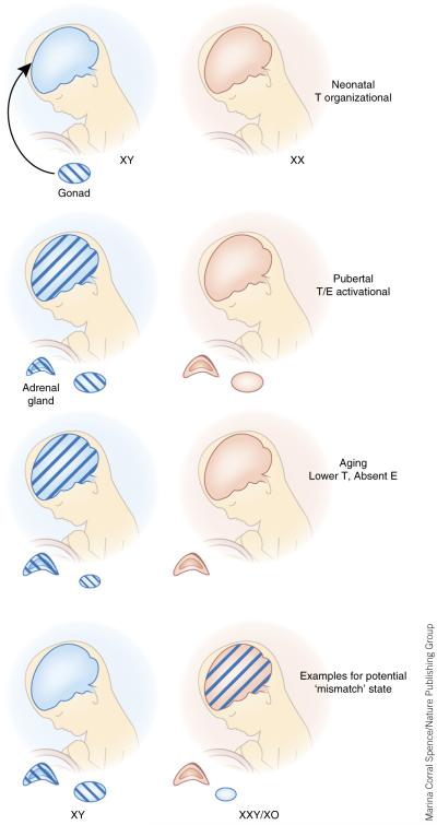
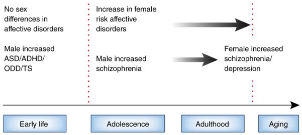
Sex differences in stress responses can be found at all stages of life and are related to both the organizational and activational effects of gonadal hormones and to genes on the sex chromosomes. As stress dysregulation is the most common feature across neuropsychiatric diseases, sex differences in how these pathways develop and mature may predict sex-specific periods of vulnerability to disruption and increased disease risk or resilience across the lifespan. The aging brain is also at risk to the effects of stress, where the rapid decline of gonadal hormones in women combined with cellular aging processes promote sex biases in stress dysregulation. In this Review, we discuss potential underlying mechanisms driving sex differences in stress responses and their relevance to disease. Although stress is involved in a much broader range of diseases than neuropsychiatric ones, we highlight here this area and its examples across the lifespan.
Figures
Comment in
-
Psychogenic nonepileptic seizures and sex differences in stress responses.Epilepsia. 2016 May;57(5):853. doi: 10.1111/epi.13350. Epilepsia. 2016. PMID: 27160800 No abstract available.
References
-
- Vale W, Spiess J, Rivier C, Rivier J. Characterization of a 41-residue ovine hypothalamic peptide that stimulates secretion of corticotropin and beta-endorphin. Science. 1981;213:1394–1397. - PubMed
-
- Rivier C, Vale W. Effects of corticotropin-releasing factor, neurohypophyseal peptides, and catecholamines on pituitary function. Fed. Proc. 1985;44:189–195. - PubMed
-
- Sawchenko PE, et al. The functional neuroanatomy of corticotropin-releasing factor. Ciba Found. Symp. 1993;172:5–21. - PubMed
-
- Kirby LG, Rice KC, Valentino RJ. Effects of corticotropin-releasing factor on neuronal activity in the serotonergic dorsal raphe nucleus. Neuropsychopharmacology. 2000;22:148–162. - PubMed
-
- Rivier C, Vale W. Modulation of stress-induced ACTH release by corticotropin-releasing factor, catecholamines and vasopressin. Nature. 1983;305:325–327. - PubMed
Publication types
MeSH terms
Grants and funding
- R01 DA037289/DA/NIDA NIH HHS/United States
- R01 AG048839/AG/NIA NIH HHS/United States
- MH091258/MH/NIMH NIH HHS/United States
- K24 DA030301/DA/NIDA NIH HHS/United States
- R33 MH104184/MH/NIMH NIH HHS/United States
- AG048839/AG/NIA NIH HHS/United States
- P50 MH099910/MH/NIMH NIH HHS/United States
- MH099910/MH/NIMH NIH HHS/United States
- R01 MH091258/MH/NIMH NIH HHS/United States
- MH087597/MH/NIMH NIH HHS/United States
- DA030301/DA/NIDA NIH HHS/United States
- MH108286/MH/NIMH NIH HHS/United States
- R01 MH087597/MH/NIMH NIH HHS/United States
- R01 MH073030/MH/NIMH NIH HHS/United States
- R21 MH104184/MH/NIMH NIH HHS/United States
- R37 MH108286/MH/NIMH NIH HHS/United States
- MH073030/MH/NIMH NIH HHS/United States
- MH104184/MH/NIMH NIH HHS/United States
LinkOut - more resources
Full Text Sources
Other Literature Sources
Medical
Research Materials
