. 2015 Nov 13;290(46):27712-22.
doi: 10.1074/jbc.M115.681205. Epub 2015 Sep 24.

Assembling a Correctly Folded and Functional Heptahelical Membrane Protein by Protein Trans-splicing

Affiliations

Assembling a Correctly Folded and Functional Heptahelical Membrane Protein by Protein Trans-splicing

Michaela Mehler et al. J Biol Chem. .

Abstract

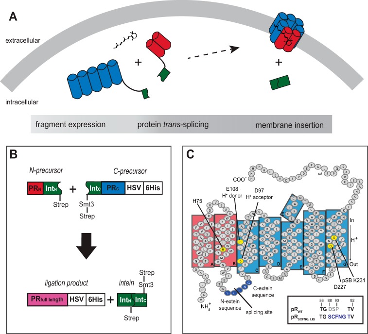
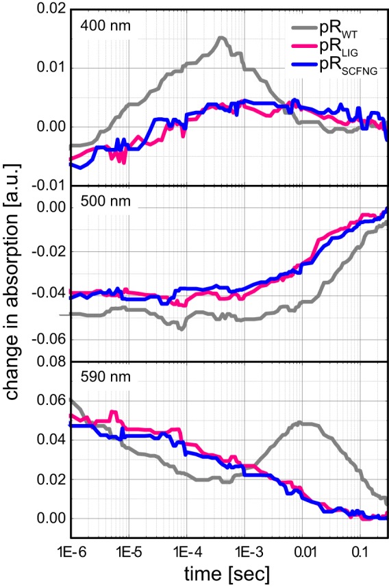
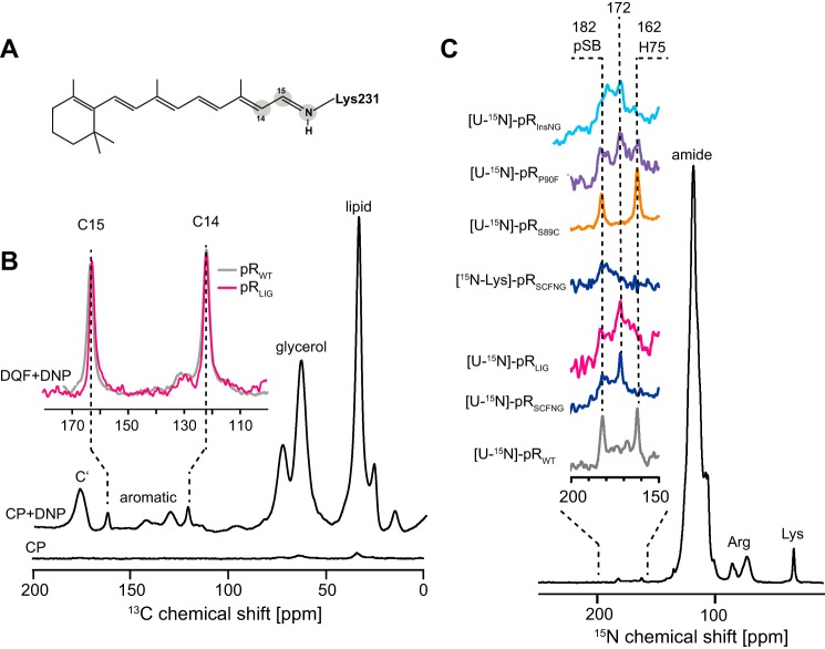
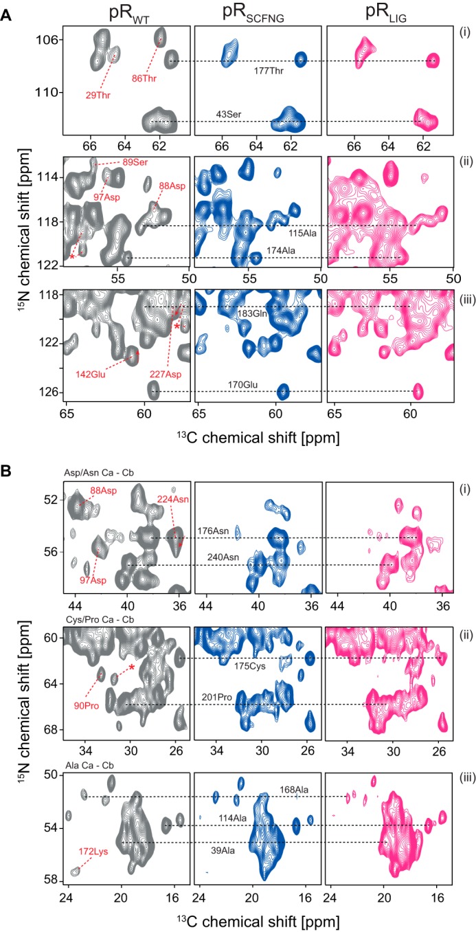
Protein trans-splicing using split inteins is well established as a useful tool for protein engineering. Here we show, for the first time, that this method can be applied to a membrane protein under native conditions. We provide compelling evidence that the heptahelical proteorhodopsin can be assembled from two separate fragments consisting of helical bundles A and B and C, D, E, F, and G via a splicing site located in the BC loop. The procedure presented here is on the basis of dual expression and ligation in vivo. Global fold, stability, and photodynamics were analyzed in detergent by CD, stationary, as well as time-resolved optical spectroscopy. The fold within lipid bilayers has been probed by high field and dynamic nuclear polarization-enhanced solid-state NMR utilizing a (13)C-labeled retinal cofactor and extensively (13)C-(15)N-labeled protein. Our data show unambiguously that the ligation product is identical to its non-ligated counterpart. Furthermore, our data highlight the effects of BC loop modifications onto the photocycle kinetics of proteorhodopsin. Our data demonstrate that a correctly folded and functionally intact protein can be produced in this artificial way. Our findings are of high relevance for a general understanding of the assembly of membrane proteins for elucidating intramolecular interactions, and they offer the possibility of developing novel labeling schemes for spectroscopic applications.

Keywords: intein; membrane protein; photoreceptor; protein folding; protein splicing; proton pump; seven-helix receptor; solid-state NMR.


Figures

FIGURE 1.
FIGURE 1.
A, in vivo protein trans-splicing of two α-helical fragments of pR on the basis of split inteins. B, design of the pR-intein segments. The N-terminal precursor contains helices A and B of pR, followed by DnaEN102 and a Strep tag. The C-terminal precursor consists of a Strep tag, an Smt3 solubility tag, NpuΔ36, and helices C, D, E, F, and G of pR, followed by an HSV-His6 tag. By protein trans-splicing, the solubility and both Strep tags are excised, resulting in full-length HSV-His6-tagged pR (pRLIG). C, topology plot of pRWT and pRLIG/pRSCFNG. To ensure an efficient splicing reaction, the BC loop amino acid sequence was modified as depicted. pSB, protonated Schiff base.
FIGURE 2.
FIGURE 2.
A–C, Western blots (A and B) and SDS-PAGE (C) of the protein trans-splicing reaction. 4 h after the first and 1 h after the second induction, the presence of N and C precursors as well as ligated protein, pRLIG, was detected in the anti-Strep (A) and anti-His6 blot (B). After 20 h, a C-terminal cleavage product (C-precursorΔSmt3) and the excised intein (inteinΔSmt3) became visible. After 24 h, precursors and pRLIG were also detectable in SDS-PAGE (C).
FIGURE 3.
FIGURE 3.
A, stationary light absorption spectrum of pRLIG at pH 7. The ratio of the absorption at 280 nm was twice as strong compared with 520 nm, demonstrating high sample purity. The values of λmax of pRSCFNG and pRLIG were nearly identical, but they were blue-shifted by 5 nm with respect to pRWT. a.u., arbitrary units. B, pH titration of max for pRLIG, pRSCFNG, pRWT, and pRInsNG in the range of pH 4–11. The pKa values of the primary proton acceptor Asp97 for each sample were determined from the inflections points of the curve. C, pH titration of λmax for pRD88S, pRS89C, and pRP90F. The pKa of Asp97 increases upon introduction of D88S and P90F but is reduced with the S89C mutation. The observation that the pKa is not altered in pRLIG and pRSCFNG might be explained by compensating effects when replacing the residue triplet Asp88-Ser89-Pro90 with Ser88-Cys89-Phe90.
FIGURE 4.
FIGURE 4.
A, CD spectra of pRWT, pRSCFNG, and pRLIG showing similarly shaped spectra, indicating a primarily α-helical fold. B, the temperature dependence of the α-helical band was followed at 222 nm. A melting temperature of 100 °C was determined for pRWT, which is slightly lower for pRSCFNG (88 °C) and pRLIG (80 °C).
FIGURE 5.
FIGURE 5.
Flash photolysis experiments on pRLIG, pRSCFNG and pRWT. The formation and decay of the M state were monitored at 400 nm, the ground-state bleach at 500 nm, and the decay of the K state and buildup of late intermediates were observed at 590 nm. pRLIG and pRSCFNG are indistinguishable from each other but show an accelerated photocycle compared with pRWT. a.u., arbitrary units.
FIGURE 6.
FIGURE 6.
A, isotope labeling of the retinal-Schiff base complex. B, DNP-enhanced 13C MAS NMR spectrum of 14,15-13C-all-trans-retinal bound to pRWT and pRLIG. The natural abundance of 13C signals was suppressed by a double quantum filter so that only signals C14 and C15 of the retinal cofactor remained. The chemical shift of resonances in pRLIG was identical to pRWT. C, 15N MAS NMR spectra of [U-15N]pRWT, pRSCFNG, and pRLIG as well as of [15N-Lys]pRSCFNG and of the mutants [U-15N]pRS89C, pRP90F, and pRInsNG. The signal of His75 was shifted from 162 ppm in pRWT to 172 ppm in pRLIG and pRSCFNG. The signal of the protonated Schiff base (pSB) was broadened but not shifted in comparison with pRWT. The spectrum of [15N-Lys]pRSCFNG confirmed the assignment of both Schiff base and His75 resonances. The spectrum of the pRS89C mutant was identical to that of pRWT. In contrast, the P90F mutant also showed a broad peak at 182 ppm and two broad peaks at 172 and 162 ppm. The spectrum of pRInsNG was very similar to the ligation product. It also showed two broad peaks at 182 and 172 ppm. The spectra in A were recorded at 400 MHz/263 GHz at 115 K. The spectra in B were acquired at 850 MHz at 275 K. The proteins were reconstituted into l-α-dimyristoylphosphatidylcholine/1,2-dimyristoyl-sn-glycero-3-phosphate lipid bilayers. The pH was adjusted to 9.
FIGURE 7.
FIGURE 7.
A and B, comparison of (A) 15N-13C (NCA) and (B) 13C-13C (proton-driven spin diffusion) correlation spectra of pRWT, pRSCFNG, and pRLIG. Resolution and line width were comparable between all three samples. Most peak positions were conserved, as shown for some selected resonances, illustrating that assembling a membrane protein via protein trans-splicing does not alter its structure. Because of the alteration in the BC loop, some resonances disappeared in pRLIG and pRSCFNG compared with pRWT (Asp88 in Aii/Bi, Ser89 in Aii, and Pro90 in Bii). A few other peaks in pRWT were shifted or broadened in pRSCFNG and pRLIG (Thr29, Thr86, Asp97, Glu142, Lys172, Asn224, and an unassigned cross peak (asterisk)). The signal-to-noise ratio for the pRLIG spectra was slightly reduced because of the lower amount of available protein. As for Fig. 6C, spectra were acquired at 850 MHz at 270 K. The proteins were reconstituted into l-α-dimyristoylphosphatidylcholine/1,2-dimyristoyl-sn-glycero-3-phosphate lipid bilayers. The pH was adjusted to 9.

References

    1. Volkmann G., and Iwaï H. (2010) Protein trans-splicing and its use in structural biology: opportunities and limitations. Mol. Biosyst. 6, 2110–2121 - PubMed
    1. Muir T. W., Sondhi D., and Cole P. A. (1998) Expressed protein ligation: a general method for protein engineering. Proc. Natl. Acad. Sci. U. S. A. 95, 6705–6710 - PMC - PubMed
    1. Al-Ali H., Ragan T. J., Gao X., and Harris T. K. (2007) Reconstitution of modular PDK1 functions on trans-splicing of the regulatory PH and catalytic kinase domains. Bioconjugate Chem. 18, 1294–1302 - PMC - PubMed
    1. Yang J.-Y., and Yang W. Y. (2009) Site-Specific Two-Color Protein Labeling for FRET Studies Using Split Inteins. J. Am. Chem. Soc. 131, 11644–11645 - PubMed
    1. Ozawa T., and Umezawa Y. (2001) Detection of protein-protein interactions in vivo based on protein splicing. Curr. Opin. Chem. Biol. 5, 578–583 - PubMed

Publication types

LinkOut - more resources