Review
. 2015 Nov;56(11):2061-9.
doi: 10.1194/jlr.R062893. Epub 2015 Sep 24.

Dermal white adipose tissue: a new component of the thermogenic response

Affiliations
Review

Dermal white adipose tissue: a new component of the thermogenic response

Caroline M Alexander et al. J Lipid Res. 2015 Nov.

Abstract

Recent literature suggests that the layer of adipocytes embedded in the skin below the dermis is far from being an inert spacer material. Instead, this layer of dermal white adipose tissue (dWAT) is a regulated lipid layer that comprises a crucial environmental defense. Among all the classes of biological molecules, lipids have the lowest thermal conductance and highest insulation potential. This property can be exploited by mammals to reduce heat loss, suppress brown adipose tissue activation, reduce the activation of thermogenic programs, and increase metabolic efficiency. Furthermore, this layer responds to bacterial challenge to provide a physical barrier and antimicrobial disinfection, and its expansion supports the growth of hair follicles and regenerating skin. In sum, this dWAT layer is a key defensive player with remarkable potential for modifying systemic metabolism, immune function, and physiology. In this review, we discuss the key literature illustrating the properties of this recently recognized adipose depot.

Keywords: adipocytes; antimicrobial; cytokines; diabetes; environmental defense; follicular development; insulation; skin; thermogenesis.


Figures

Fig. 1.
Fig. 1.
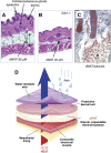
dWAT in skin from mice and men. A: Morphology of mouse adult skin. Hematoxylin and eosin-stained section of belly skin from 11-week-old BALB/cJ female, housed at 21°C. B: Sample from corresponding Sdc1−/− mouse showing deficient dWAT. C: Human fetal skin (Azan-stained section, 30 μm) from the facial region at 19.5 weeks gestation showing the appearance of a cluster of adipose lobules very close to the base of a developing hair follicle. D: Diagram of the lamellar structure of skin. The multi-layered structure of skin is drawn by analogy to “Gore-Tex” to emphasize the different roles of each layer. Specifically, this illustration emphasizes the role that dWAT plays in the reduction of heat loss from a mammalian core body temperature (approximately 37°C) to the variable environmental temperature.
Fig. 2.
Fig. 2.
Regulation of dWAT. Three separate regulators of dWAT expansion are indicated in red. This diagram indicates a cross-section of an average mammal, coated in skin with a subjacent layer of dWAT, expanded or not (adipocytes are shown as hexagons). The physiology of skin determines the physiology of all internal organs. dWAT expands in response to cold exposure to provide insulation, and in response to bacterial infection, where it counters microbial colonization, and in response to the hair follicle cycle, to support follicular invagination. Together, these responses comprise a comprehensive defensive strategy for the mammalian ectoderm.
Fig. 3.
Fig. 3.
Insulating sleeve of dWAT. A: dWAT was visualized (and quantified) in 3D using high resolution MRI (fat only) for an adult female BALB/cJ mouse. B. Typical adult mouse skin stained with hematoxylin and eosin to show the patched asynchronous pattern of anagen. I. Kasza et al., unpublished.
Fig. 4.
Fig. 4.
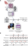
The activation of thermogenic defenses. A: The integration of adipocyte depots that provide thermogenic homeostatis is diagrammed. Perceived body temperature is shown as a line in the center of the diagram. When body temperature drops, cold sensors are activated (including hypothalamic, cardiac, and perhaps local sources; shown here are macrophages in WAT tissues; see text for details). Effectors induce the activation of facultative thermogenic depots (including BAT and brite depots) that become lipolytic, generating heat (shown as pink wiggle lines) from uncoupled mitochondria and lipids to fuel the β-oxidation required for warming. The pattern of BAT activation, revealed by PET imaging, is shown for a human subject [reproduced from (74), with permission]. As the temperature challenge is remediated, thermogenesis is deactivated. The timeline for BAT activation in response to a (noxious) 4°C challenge is quick (less than 30 minutes) (28); otherwise, the periodicity of this cycle is not known. The efficiency of heat retention is determined by the total insulation, in part determined by the dWAT layer. This dWAT layer acts as a third component of this circuit, and responds to overall ambient temperature, but slowly (days). B: Hypothetical patterns of thermogenic activation. We propose that the overall time spent with thermal defenses activated is a function of the absolute temperature challenge, the efficiency of remediation with activation of thermal defenses, and the level of insulation. The body temperature of mice is shown as a black line and the activation of thermal defenses is shown above as a red line. A typical pattern for mice housed at 20–25°C is compared with those moved to 4°C (extreme cold) and the complete absence of thermogenesis observed under thermoneutral conditions. As a comparison, the lack of insulation in dWAT-deficient mice may slow remediation of cooling body temperature and activate the cycle more frequently, potentially leading to chronic activation. In contrast, high levels of dWAT observed in obese mice (and perhaps also in obese humans) leads to a hyper-insulated phenotype and little activation of thermogenesis.
Fig. 5.
Fig. 5.
The under-insulated phenotype. A: A diagrammatic representation of the cross-section of a mammal (as for Fig. 2) coated in skin and protected from heat loss (pink arrow) by a layer of dWAT. B: When dWAT is deficient, the thermogenic program is chronically activated, leading to systemic hyper-activation of key metabolic checkpoints, such as p38α. Symptoms of under-insulation include chronic WAT/brite/BAT activation at cool housing temperatures (room temperature for mice), depleted liver glycogen, and susceptibility to torpor in response to fasting. Note that total energy expenditure may not be increased in under-insulated mice; for example, energy expenditure was not affected in Sdc1−/− mice (6), and indeed the lack of response of the adipostat to thermogenic load has been discussed before (4). Therefore, this is not considered a core component of this phenotype.

References

    1. Sanchez-Gurmaches J., Guertin D. A. 2014. Adipocyte lineages: tracing back the origins of fat. Biochim. Biophys. Acta. 1842: 340–351. - PMC - PubMed
    1. Fried S. K., Lee M. J., Karastergiou K. 2015. Shaping fat distribution: New insights into the molecular determinants of depot- and sex-dependent adipose biology. Obesity (Silver Spring). 23: 1345–1352. - PMC - PubMed
    1. Gesta S., Tseng Y. H., Kahn C. R. 2007. Developmental origin of fat: tracking obesity to its source. Cell. 131: 242–256. - PubMed
    1. Nedergaard J., Cannon B. 2014. The browning of white adipose tissue: some burning issues. Cell Metab. 20: 396–407. - PubMed
    1. Sun K., Kusminski C. M., Scherer P. E. 2011. Adipose tissue remodeling and obesity. J. Clin. Invest. 121: 2094–2101. - PMC - PubMed

Publication types