. 2015 Dec;88(6):1045-54.
doi: 10.1124/mol.115.101451. Epub 2015 Sep 24.

A Novel Hsp90 Inhibitor Activates Compensatory Heat Shock Protein Responses and Autophagy and Alleviates Mutant A53T α-Synuclein Toxicity

Affiliations

A Novel Hsp90 Inhibitor Activates Compensatory Heat Shock Protein Responses and Autophagy and Alleviates Mutant A53T α-Synuclein Toxicity

Rui Xiong et al. Mol Pharmacol. 2015 Dec.

Abstract

A potential cause of neurodegenerative diseases, including Parkinson's disease (PD), is protein misfolding and aggregation that in turn leads to neurotoxicity. Targeting Hsp90 is an attractive strategy to halt neurodegenerative diseases, and benzoquinone ansamycin (BQA) Hsp90 inhibitors such as geldanamycin (GA) and 17-(allylamino)-17-demethoxygeldanamycin have been shown to be beneficial in mutant A53T α-synuclein PD models. However, current BQA inhibitors result in off-target toxicities via redox cycling and/or arylation of nucleophiles at the C19 position. We developed novel 19-substituted BQA (19BQA) as a means to prevent arylation. In this study, our data demonstrated that 19-phenyl-GA, a lead 19BQA in the GA series, was redox stable and exhibited little toxicity relative to its parent quinone GA in human dopaminergic SH-SY5Y cells as examined by oxygen consumption, trypan blue, 3-(4,5-dimethylthiazol-2-yl)-2,5-diphenyl-tetrazolium bromide (MTT), and apoptosis assays. Meanwhile, 19-phenyl-GA retained the ability to induce autophagy and potentially protective heat shock proteins (HSPs) such as Hsp70 and Hsp27. We found that transduction of A53T, but not wild type (WT) α-synuclein, induced toxicity in SH-SY5Y cells. 19-Phenyl-GA decreased oligomer formation and toxicity of A53T α-synuclein in transduced cells. Mechanistic studies indicated that mammalian target of rapamycin (mTOR)/p70 ribosomal S6 kinase signaling was activated by A53T but not WT α-synuclein, and 19-phenyl-GA decreased mTOR activation that may be associated with A53T α-synuclein toxicity. In summary, our results indicate that 19BQAs such as 19-phenyl-GA may provide a means to modulate protein-handling systems including HSPs and autophagy, thereby reducing the aggregation and toxicity of proteins such as mutant A53T α-synuclein.


Figures

Fig. 1.
Fig. 1.
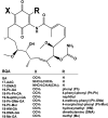
Chemical structures of BQAs. Substitutions at the 19-position (R) include phenyl (Ph), 4-phenyl-phenyl (Ph-Ph), naphthyl, 4-methoxy-phenyl (Ph-OMe), 4-morpholinyl-phenyl (Ph-Mor), hydroxymethyl (HM), dimethylamino (DMA), and methyl (Me).
Fig. 2.
Fig. 2.
19BQAs exerted decreased toxicity compared with their parent quinone GA. (A) Growth inhibition induced by 19BQAs in 5Y cells. Growth inhibition was measured using the MTT assay. Cells were treated with BQAs for 4 hours, then allowed to grow for an additional 72 hours. (B) Cells were treated with GA and 19-Ph-GA at 0.25 μM for 24 hours and harvested for determination of cell number using a trypan blue exclusion assay. (C) Apoptosis was measured using annexin V/PI cell staining in combination with flow cytometry in cells after exposure to the equivalent concentration (5 μM) of GA or 19-Ph-GA for 16 hours. (D) Oxygen consumption was measured in cells in the absence (basal) or presence of the indicated compounds. Measurements were made over 10 minutes using a Clark electrode at 37°C. Menadione was included as a redox-cycling quinone (positive control). Results are expressed as the mean ± S.D., n = 3. *P < 0.05, ***P < 0.001 is considered significantly different from basal by one-way analysis of variance using Dunnett’s multiple comparison test. 19-DMA-GA, 19-dimethylamino-GA; 19-HM-GA, 19-hydroxymethyl-GA; 19-Me-GA, 19-methyl-GA; 19-Ph-Mor-GA, 19-(4-morpholinyl-phenyl)-GA; 19-Ph-OMe-GA, 19-(4-methoxy-phenyl)-GA; 19-Ph-Ph-GA, 19-(4-phenyl-phenyl)-GA.
Fig. 3.
Fig. 3.
Treatment with 19BQAs induced HSPs in 5Y cells. Immunoblot analysis of biomarkers of Hsp90 inhibition following treatment with BQAs at the indicated concentrations for 16 hours. Note that many 19BQAs induced potent expression of Hsp70 and Hsp27. 19-DMA-GA, 19-dimethylamino-GA; 19-HM-GA, 19-hydroxymethyl-GA; 19-Me-GA, 19-methyl-GA; 19-Ph-Mor-GA, 19-(4-morpholinyl-phenyl)-GA; p-Akt, phosphorylated Akt; 19-Ph-OMe-GA, 19-(4-methoxy-phenyl)-GA; 19-Ph-Ph-GA, 19-(4-phenyl-phenyl)-GA.
Fig. 4.
Fig. 4.
Overexpression of human mutant A53T α-Syn induced protein handling changes and toxicity in 5Y cells. (A) One day after seeding, cells were transduced with mutant A53T α-Syn (MOI from 100 to 800 plaque-forming units/cell) and WT α-Syn (MOI 400 plaque-forming units/cell) for 72 hours. Cell viability was then determined by the MTT assay. (B) Overexpression of A53T α-Syn inhibited intracellular 20/26S proteasomal activity (chymotrypsin-like active site) in a dose-dependent manner. (C) Hsp70 and Hsp27 protein levels and induction of the ER stress response and autophagy following transduction with A53T α-Syn. (D) MOI-dependent increase in higher molecular weight α-Syn oligomers was detected 72 hours after transduction with A53T α-Syn, but not WT α-Syn, and green fluorescent protein (GFP) adenovirus vector control (MOI 400 plaque-forming units/cell). β-Actin was included as a loading control. A representative blot from three separate experiments is shown for each figure. Values in (A) and (B) are presented as the mean ± S.D. (n = 3); *P < 0.05 is considered significant to control group (one-way analysis of variance using Dunnett’s multiple comparison test). P-eIF2α, phosphorylated eIF2α; MOI, multiplicity of infection.
Fig. 5.
Fig. 5.
19-Ph-GA significantly attenuated A53T α-Syn–induced toxicity in 5Y cells. (A) Treatment with low concentrations of 19-Ph-GA (0.5–1 μM) for 72 hours was not cytotoxic to 5Y cells. (B) After 24-hour incubation with adenovirus expressing mutant A53T α-Syn (400 plaque-forming units/cell), cells were exposed to the indicated concentrations of 19-Ph-GA for 48 hours, after which cell viability was determined by the MTT assay. Triplicate treatments in 48-well plates were used. Results represent one experiment typical of n = 5. Values are presented as the mean ± S.D. (n = 3); *P < 0.05, **P < 0.01 is considered significant compared with control in (A) and relative to the A53T α-Syn group in (B), respectively, by one-way analysis of variance using Tukey’s multiple comparison test.
Fig. 6.
Fig. 6.
19-Ph-GA significantly reduced A53T α-Syn oligomer formation through induction of heat shock protein responses and autophagy. (A and B) α-Syn oligomers (denatured) were significantly reduced after 48-hour post-treatment of 5Y cells with 19-Ph-GA. (B) Fold changes of α-Syn oligomers in (A) after the drug treatment are normalized to respective controls and estimated by densitometry. (C) Exposure of 5Y cells to 19-Ph-GA resulted in decreased levels of native high molecular weight α-Syn oligomers as well as an increase in the smaller (76 kDa) oligomer species. (D) After 48-hour post-treatment with 19-Ph-GA, Hsp70 and Hsp27 protein levels were elevated 2- to 3-fold, but biomarkers of autophagy and ER stress were not significantly perturbed. (E) Time-course study showed that 19-Ph-GA was able to upregulate both HSPs and LC3 II expression in 5Y cells. Note that exposure to 19-Ph-GA for 24 hours induced the maximum increase in LC3 II, suggesting 19-Ph-GA stimulated a temporal autophagic flux in 5Y cells. (F and G) Significant induction of HSPs and LC3 II could be detected in A53T α-Syn–overexpressing cells after post-treatment with 19-Ph-GA (0.5 μM) for 24 hours, suggesting that HSP response and autophagic flux were stimulated by 19-Ph-GA at relatively early time points. β-Actin was included as a loading control. A representative blot from three separate experiments is shown for each figure. (G) Fold changes of Hsp70, Hsp27, and LC3 II in (F) after the drug treatment at the indicated times are normalized to respective controls and estimated by densitometry. Values in (B) and (G) are presented as the mean ± S.D. (n = 3); *P < 0.05 considered significant compared with the A53T α-Syn group by one-way analysis of variance using Tukey’s multiple comparison test. p-eIF2α, phosphorylated eIF2α.
Fig. 7.
Fig. 7.
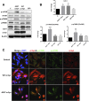
mTOR/p70S6K signaling was activated by overexpression of A53T but not WT α-Syn. (A) Overexpression of A53T, but not WT for 72h, α-Syn significantly increased the levels of phosphorylated mTOR and p70S6K in 5Y cells. An increased amount of LC3 II was observed in both A53T and WT α-Syn–overexpressing cells, suggesting that autophagy was not directly mediated via the mTOR/p70S6K signaling. The white dividing line in the LC3 II blot indicates omission of three lanes on the gel; all lanes shown were from the same gel. (B) Fold changes of LC3 II, p-mTOR, and p-p70S6K levels in (A) are normalized to respective controls and estimated by densitometry. These values are presented as the mean ± S.D. (n = 3); *P < 0.05 is considered significant relative to the control group using Dunnett’s multiple comparison test. (C) Increased expression of p-mTOR was observed in A53T, but not WT, α-syn–overexpressing cells. After transduction with adenovirus expressing A53T α-Syn or WT α-Syn at MOI 400 plaque-forming units/cell for 72 hours, 5Y cells were fixed and immunostained for α-Syn (red), p-mTOR (green), and nuclei (DAPI, blue) and then examined by confocal microscopy. Note that the colocalization of p-mTOR and α-Syn (white arrow), suggesting an interaction between p-mTOR and A53T α-Syn, may exist. Ad, adenovirus; GFP, green fluorescent protein; DAPI, 4′,6-diamidino-2-phenylindole; MOI, multiplicity of infection.
Fig. 8.
Fig. 8.
19-Ph-GA decreased mTOR activation that was associated with A53T α-Syn–induced toxicity. (A) Post-treatment with 19-Ph-GA for 48 hours significantly blocked the activation of p-mTOR and p-p70S6K induced by A53T α-Syn. (B) Fold changes of LC3 II, p-mTOR, and p-p70S6K levels in (A) are normalized to respective controls and estimated by densitometry. These values are presented as the mean ± S.D. (n = 3); *P < 0.05 is considered significant relative to the control group; #P < 0.05 is considered significant compared with the A53T α-Syn group by one-way analysis of variance using Tukey’s multiple comparison test.
Fig. 9.
Fig. 9.
Protective effects of 19-Ph-GA on A53T α-Syn induced protein handling changes and toxicity in 5Y cells. Overexpression of A53T α-Syn (labeled in red) resulted in α-Syn oligomer formation and toxicity via perturbing major protein handling systems, including increased proteasomal inhibition, ER stress response, and decreased levels of HSPs. Autophagy could be activated by overexpression of either A53T or WT α-Syn (labeled in black), excluding the possibility that autophagic cell death was induced specifically by A53T α-Syn overexpression. mTOR/p70S6K signaling was not directly involved in A53T α-Syn–induced autophagy but was positively related to the toxicity of A53T α-Syn. 19-Ph-GA (labeled in blue) activated Hsp70, Hsp27, and autophagy and ameliorated A53T α-Syn–induced mTOR activation. Collectively, these mechanisms may contribute to protection against A53T α-Syn oligomer formation and toxicity.

Similar articles

Cited by

References

    1. Auluck PK, Chan HYE, Trojanowski JQ, Lee VM-Y, Bonini NM. (2002) Chaperone suppression of alpha-synuclein toxicity in a Drosophila model for Parkinson’s disease. Science 295:865–868. - PubMed
    1. Baldo B, Weiss A, Parker CN, Bibel M, Paganetti P, Kaupmann K. (2012) A screen for enhancers of clearance identifies huntingtin as a heat shock protein 90 (Hsp90) client protein. J Biol Chem 287:1406–1414. - PMC - PubMed
    1. Blair LJ, Sabbagh JJ, Dickey CA. (2014) Targeting Hsp90 and its co-chaperones to treat Alzheimer’s disease. Expert Opin Ther Targets 18:1219–1232. - PMC - PubMed
    1. Bové J, Martínez-Vicente M, Vila M. (2011) Fighting neurodegeneration with rapamycin: mechanistic insights. Nat Rev Neurosci 12:437–452. - PubMed
    1. Chang C-H, Drechsel DA, Kitson RRA, Siegel D, You Q, Backos DS, Ju C, Moody CJ, Ross D. (2014) 19-substituted benzoquinone ansamycin heat shock protein-90 inhibitors: biological activity and decreased off-target toxicity. Mol Pharmacol 85:849–857. - PMC - PubMed

Publication types

MeSH terms