. 2015 Oct 16;350(6258):328-34.
doi: 10.1126/science.aad0395. Epub 2015 Sep 24.

RIPK1 and NF-κB signaling in dying cells determines cross-priming of CD8⁺ T cells

Affiliations

RIPK1 and NF-κB signaling in dying cells determines cross-priming of CD8⁺ T cells

Nader Yatim et al. Science. .

Abstract

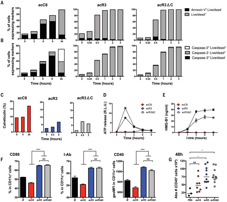
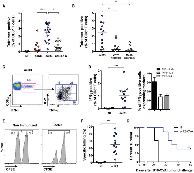
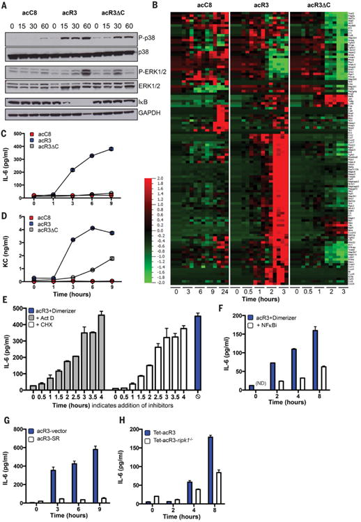
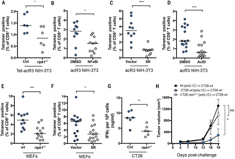
Dying cells initiate adaptive immunity by providing both antigens and inflammatory stimuli for dendritic cells, which in turn activate CD8(+) T cells through a process called antigen cross-priming. To define how different forms of programmed cell death influence immunity, we established models of necroptosis and apoptosis, in which dying cells are generated by receptor-interacting protein kinase-3 and caspase-8 dimerization, respectively. We found that the release of inflammatory mediators, such as damage-associated molecular patterns, by dying cells was not sufficient for CD8(+) T cell cross-priming. Instead, robust cross-priming required receptor-interacting protein kinase-1 (RIPK1) signaling and nuclear factor κB (NF-κB)-induced transcription within dying cells. Decoupling NF-κB signaling from necroptosis or inflammatory apoptosis reduced priming efficiency and tumor immunity. Our results reveal that coordinated inflammatory and cell death signaling pathways within dying cells orchestrate adaptive immunity.


Figures

Fig. 1
Fig. 1. Necroptotic cells release DAMPs and induce dendritic cell maturation
(A to C) NIH-3T3 cells expressing the death constructs were stimulated with dimerizer and cells harvested at the indicated time points and stained with Annexin-V and Live/Dead reagent (A); cleaved caspase-3 antibody and Live/Dead reagent (B); or calreticulin antibody (C). Cells that are Annexin V+ Live/Dead (indicating phosphatidylserine exposure prior to membrane permeabilization) or cleaved-caspase-3+ (indicating the activation of executionner caspases) are undergoing apopotsis. At later time points (24hrs), staining with Live/Dead reagent indicates loss of plasma membrane integrity and characterizes secondary necrotic cells. Rapid membrane permeabilzation without activation of executionner caspases (Live-Dead+ Caspase-3-) is a feature of necroptosis. N=2, results represent one representative experiment (D and E) ATP and HMGB1 were quantified from dying cell culture supernatants. N=3, results are reported as mean +/− SEM of triplicates of one representative experiment. (F) BMDCs were co-cultured with dimerizer treated acC8-, acR3- and acR3ΔC-expressing cells for 24h and DC maturation phenotype was assessed by flow cytometry. N=4, results are reported as mean +/− SEM of triplicates of one representative experiment. (G) 2×10⁶ dimerizer treated cells were injected into the peritoneal cavity of WT C57BL/6 mice. 48h later, peritoneal cells were collected and immune cells were enumerated by cytometry. N=2, bars indicate mean of two pooled independent experiment with 4-5 mice per group (except PBS group). Each circle represent one mouse. p values were determined using one-way ANOVA test for (F) and Kruskal-Wallis test (KW - multigroup comparision) followed by a Dunn's post-test, comparing each group to PBS group for (G). *P < 0.05; ****P < 0.0001. acC8 = Casp8 apoptosis; acR3 = RIPK3 necroptosis; acR3ΔC = RIPK3RHIMless necroptosis; ATP= Adenosine triphosphate; HMGB1= High mobility group box 1; PBS= Phospahte-buffered saline.
Fig. 2
Fig. 2
Necroptotic cells are immunogenic and require RHIM-dependent ripoptosome formation for efficient cross-priming of CD8+ T cells. (A to G) To elicit cross-priming we intradermally injected (i.d.) OVA-expressing dying cells (H-2q) into mice (H-2b), and analyzed on day 9 post-immunization (p.i.). (A and B) Using Kb-SIINFEKL-tetramers, OVA-specific CD8+ T cells were quantified and plotted as a percentage of total CD8+ T cells. N=4 (A), N=3 (B) Bars indicate median and results are pooled from three independent experiments with 3-6 mice per group (each circle represent one mouse) (C and D) IFN-γ, TNF-α and IL-2 production in response to ex-vivo SIINFEKL peptide re-stimulation was determined. Representative FACS plots are shown and numbers indicate the percentage of gated cells (C); and frequency of IFN-γ expressing and polyfunctional cells are plotted (D) N=3, results are pooled from three independent experiments with 3-6 mice per group and reported as individual mice (each circle represent one mouse) and bars indicate median (IFN-γ) or as histogram and mean+/− SEM (TNFα and IL-2). (E and F) In vivo cytotoxicity assay was performed in acR3-OVA immunized mice. At day 8 p.i., mice were adoptively transfered with CFSE-labeled splenocytes and the frequency of CSFEhi (irrelevant peptide control) and CFSElow (SIINFEKL loaded) splenocytes (injected at a 1:1 ratio) was determined at day 9. Representative FACS plots are shown (E) and the percent of specific killing plotted (F). N=3, Bars indicate median and results are pooled from three independent experiments with 4 mice per group (each circle represent one mouse). (G) Tumor challenge experiments were performed, injecting 5×10⁵ B16F10-OVA cells on day 12 p.i., N=2, and results are reported as a survival curve from one representative experiment with 8-11 mice per group ; OVA = ovalbumin. p values were determined using KW test followed by Dunn's post-test for (A and B), Mann-Witney (MW) test for (D and F) and mice survival (G) was compared by log-rank test. *P < 0.05; **P < 0.01; ***P < 0.001; ****P < 0.0001.
Fig. 3
Fig. 3. RIPK3 oligomerization results in RIPK1-dependent activation of NF-κB mediated gene expression
(A to D), acC8-, acR3- and acR3ΔC-expressing NIH-3T3 cells were treated with dimerizer, and at indicated time points: protein extracts were analyzed by Western blot, N=2 (A); RNA was extracted for transcriptional profiling, N=3 (B); or culture supernatants were collected for luminex analysis (C and D). (E to H) IL-6 release upon addition of dimerizer was determined, after treating the cells with (E), or (F). In (E), 2μg/ml actinomycin D (Act D) or 2.5μg/ml cycloheximide (CHX) were added at the indicated time points after addition of dimerizer (t=0), and IL-6 was measured at 6h. In (F), cells were pre-teated with 10μM Wedelolactone (NFKBi) and IL-6 measured at the indicated time points. In (G), acR3 cells stably expressing control vector (acR3-vector) or mutant super-repressor IκB (acR3-SR) were used. In (H), control NIH-3T3 cells (Tet-acR3), and cells lacking RIPK1 and expressing RIPK3 2xFv under a tetracycline promoter (Tet-acR3 ripk1−/−), were treated overnight with 500ng/ml of doxycycline (Dox) before addition of dimerizer. In (C to H), N≥2, and data are presented as mean +/− SEM of triplicates from one representative experiment. Heat map indicates the relative expression of the indicated transcript (red indicating high levels and green indicating low levels of expression).
Fig. 4
Fig. 4. RIPK1 expression and NF-κB activation during cell death are required for efficient cross-priming and anti-tumor immunity
In (A) mice were immunized with Tet-acR3-OVA and Tet-acR3-OVA ripk1−/− NIH-3T3 cells. Data represent one experiment with six mice per group, bars indicate median. In (B) acR3-OVA NIH-3T3 cells were pre-treated with DMSO or BAY 11-7085 (NFκBi-10μM) for 10 min prior to addition of dimerizer and immunization; In (C) mice were immunized with acR3-OVA cells expressing NF-κB-SR or control vector. In (D) acR3-OVA were pre-treated with DMSO or ActD for 45 min prior to immunization. (E and F) OVA-expressing MEFs were transfected with 10μg/ml poly I:C and after 6h used for immunization. WT or cells lacking ripk1−/− were used in (E); or cells expressing control vector or NFκB-SR were used in (F). Cross-priming was assessed on day 9 p.i. In (B to F), N=3 and results shown are pooled from three independent experiments with 3 to 6 mice per group. (G and H) CT26 Ctrl or a CRISPR/cas9-modified line that lacks RIPK1 expression (CT26 ripk1−/−) were pIC-transfected and injected into Balb/cByJ mice. 7 days later, spleen and lymph nodes were harvested and IFNγ production was quantified (G) or mice were challenged with 5×10⁵ WT CT26 injected in the opposite flank and tumor growth was monitored every 3 days (H). N=3 and results are from one representative experiment with 6 mice per group. p values were determined using MW test (A to G) or 2-way ANOVA test (multipe group comaprison) comparing each group to nonimmunized group (NI) (J). *P < 0.05; **P < 0.01; ***P < 0.001; ****P < 0.0001.

Comment in

References

    1. Albert ML, Sauter B, Bhardwaj N. Dendritic cells acquire antigen from apoptotic cells and induce class I-restricted CTLs. Nature. 1998;392:86–89. doi: 10.1038/32183. Medline. - DOI - PubMed
    1. Albert ML, Darnell JC, Bender A, Francisco LM, Bhardwaj N, Darnell RB. Tumor-specific killer cells in paraneoplastic cerebellar degeneration. Nat Med. 1998;4:1321–1324. doi: 10.1038/3315. Medline. - DOI - PubMed
    1. Bevan MJ. Cross-priming for a secondary cytotoxic response to minor H antigens with H-2 congenic cells which do not cross-react in the cytotoxic assay. J Exp Med. 1976;143:1283–1288. doi: 10.1084/jem.143.5.1283. Medline. - DOI - PMC - PubMed
    1. Turley S, Poirot L, Hattori M, Benoist C, Mathis D. Physiological beta cell death triggers priming of self-reactive T cells by dendritic cells in a type-1 diabetes model. J Exp Med. 2003;198:1527–1537. doi: 10.1084/jem.20030966. Medline. - DOI - PMC - PubMed
    1. Iyer SS, Pulskens WP, Sadler JJ, Butter LM, Teske GJ, Ulland TK, Eisenbarth SC, Florquin S, Flavell RA, Leemans JC, Sutterwala FS. Necrotic cells trigger a sterile inflammatory response through the Nlrp3 inflammasome. Proc Natl Acad Sci USA. 2009;106:20388–20393. Medline. - PMC - PubMed

Publication types