. 2015 Oct 16;350(6258):aab4077.
doi: 10.1126/science.aab4077. Epub 2015 Sep 24.

Remote control of therapeutic T cells through a small molecule-gated chimeric receptor

Affiliations

Remote control of therapeutic T cells through a small molecule-gated chimeric receptor

Chia-Yung Wu et al. Science. .

Abstract

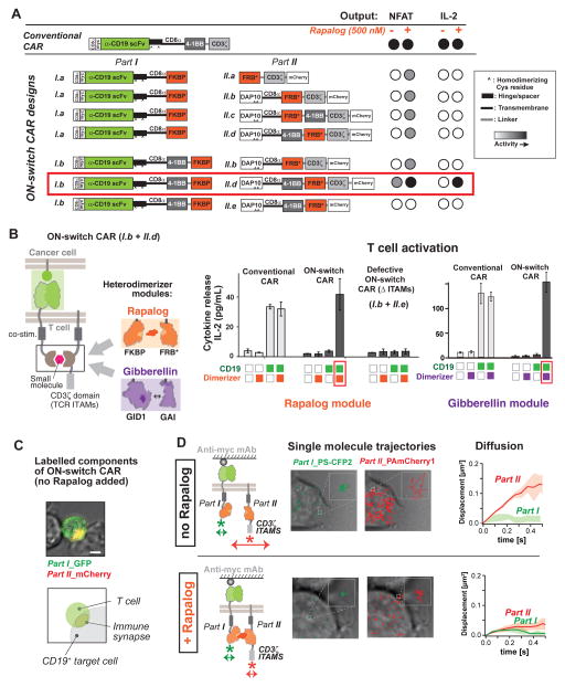
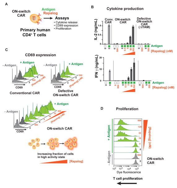
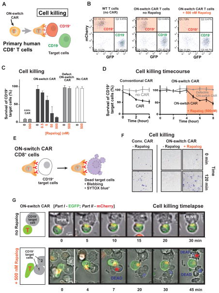
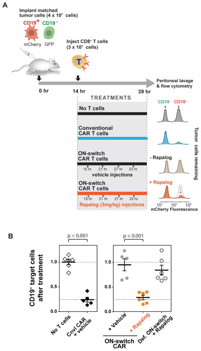
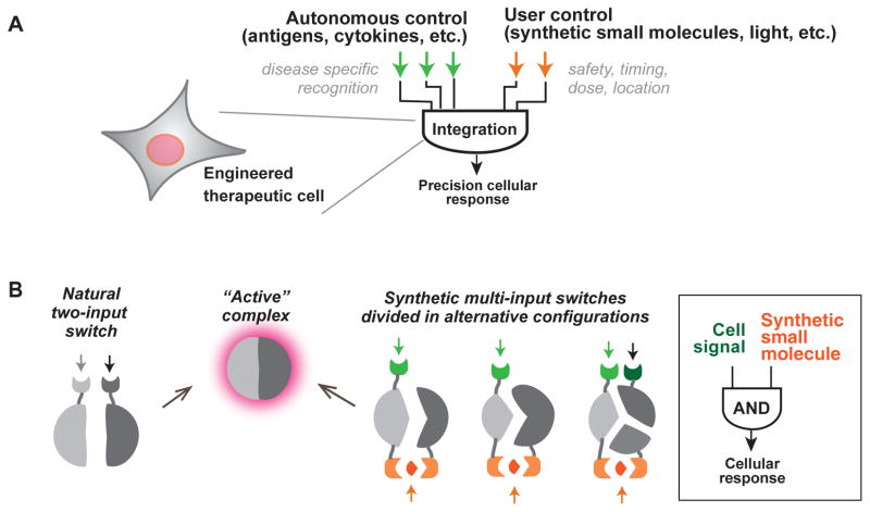
There is growing interest in using engineered cells as therapeutic agents. For example, synthetic chimeric antigen receptors (CARs) can redirect T cells to recognize and eliminate tumor cells expressing specific antigens. Despite promising clinical results, these engineered T cells can exhibit excessive activity that is difficult to control and can cause severe toxicity. We designed "ON-switch" CARs that enable small-molecule control over T cell therapeutic functions while still retaining antigen specificity. In these split receptors, antigen-binding and intracellular signaling components assemble only in the presence of a heterodimerizing small molecule. This titratable pharmacologic regulation could allow physicians to precisely control the timing, location, and dosage of T cell activity, thereby mitigating toxicity. This work illustrates the potential of combining cellular engineering with orthogonal chemical tools to yield safer therapeutic cells that tightly integrate cell-autonomous recognition and user control.


Figures

Fig. 1
Fig. 1. Strategy for design of a combinatorially activated chimeric antigen receptor (CAR) with re-arranged key signaling modules
(A) Engineered T cells expressing CARs can have both therapeutic and adverse effects. (B) User-controlled switches. A suicide switch triggers apoptosis of the engineered cells. An alternative, complementary approach is keeping the cells inactive until addition of an activating small molecule drug signal. Such an ON-switch could allow for titratable control (dialing up or down) of T cell activity. (C) Molecular strategies to control T cell activation. The normal T cell activation pathway (left) entails dual activation of the T Cell Receptor (TCR) and a co-stimulatory receptor to trigger key cellular responses such as cytokine production and proliferation. The conventional CAR (middle) combines an antigen recognition domain (single chain variable fragment; scFv) with main signaling motifs (such as ITAMs from TCR subunit CD3zeta) and co-stimulatory motifs constitutively linked in a single molecule. A strategy for constructing an ON-switch CAR utilizing a split construct (right). The split CAR design distributes key components from the conventional CAR into two physically separate polypeptides that can be conditionally re-assembled when a heterodimerizing small molecule agent is present. The design resembles an AND logic gate that requires “antigen + small molecule” combinatorial inputs for T cell activation.
Fig. 2
Fig. 2. Construction and screening of ON-switch chimeric antigen receptor (CAR) that is dependent on presence of small molecule dimerizer
(A) ON-switch CAR candidate constructs and their functional behavior. Candidate construct pairs were expressed in Jurkat T cells. Cells were incubated with K562 target cells expressing the cognate antigen CD19+ in the presence or absence of 500 nM rapalog. Activation was quantified via expression of an NFAT-dependent GFP reporter gene and production of the cytokine IL-2. The part I constructs of the ON-switch CAR share many features with the conventional CAR: the CD8α signal sequence, a Myc epitope, the anti-CD19 scFv, the CD8α hinge and transmembrane domain, in addition to the FKBP domain for heterodimerization. The part II constructs consisted of the T cell receptor CD3ζ signaling chain that is critical for T cell activation, the FRB* domain for heterodimerization, and the mCherry tag. More advanced part II variants contained the additional DAP10 ectodomain for homodimerization and the CD8α transmembrane domain for membrane anchoring. The 4-1BB costimulatory motif was inserted in various locations, depending on the construct. The best ON-switch construct (I.b + II.d) is outlined in red. (B) Response of ON-switch CAR (I.b + II.d) to rapalog and antigen stimulation. Jurkat cells expressing the specified CARs were incubated with K562 target cells expressing either the cognate antigen (CD19; green squares) or a non-cognate antigen (mesothelin; white squares). Presence of 500 nM rapalog in the sample was indicated by orange squares. Production of IL-2 after an over-night incubation was quantified by ELISA. n = 3, error bar = standard deviation. Similar results were observed for ON-switch CARs in which the rapalog heterodimerization module was replaced by an alternative module, the gibberellic acid heterodimerization module (utilizing Arabidopsis GID1 and GAI domains). The ON-switch CAR with gibberellic acid (GA) dimerizing domains requires both cognate antigen and GA (purple squares) to trigger cytokine production. (C) The ON-switch CAR components co-localize in the absence of dimerizing rapalog. Parts I and II of the receptor are labeled with GFP and mCherry, respectively. The confocal microscopy images are pseudo-colored to indicate localization of both parts. Image shows a primary human CD8+ T cell expressing the anti-CD19 ON-switch CAR engaged with a CD19+ K562 target cell in the absence of rapalog. Scale bar = 5μm. (D) Two-color single-molecule tracking shows independent movement of ON-switch CAR components in the absence of rapalog. Left panels: Jurkat T cells were adhered to a cover slip coated with an antibody to the Myc epitope in order to immobilize the receptors (extracellular region of part I CAR is tagged with Myc). Individual parts of the ON-switch CAR were each tagged with photoactivatable fluorescent proteins PS-CFP2 and PAmCherry1. Middle panels: The single molecule trajectories of part I (green) and part II (red) are superimposed on transmitted light images of the cells (gray). Right panels: The average mean-square displacement of trajectories quantifies the diffusive behavior (Solid lines: average from multiple cells. Colored band: standard deviation to represent cell-cell variability). Part I molecules are immobile due to antibody tethering, whereas in the absence of rapalog (top) part II molecules exhibit fast diffusion. In the presence of 500 nM rapalog (bottom), however, the part II molecules became immobile, confirming rapalog-induced assembly of the two-component receptor.
Fig. 3
Fig. 3. Small molecule-titratable activation of primary human helper T cell populations engineered with ON-switch CAR
(A) CD4+ T cells were purified from the peripheral blood of anonymous healthy donors, expanded, engineered with lentivirus to express CARs, and evaluated by functional assays. T cells with comparable CAR expression levels were used. The cognate antigen CD19 was presented to T cells as a cell surface protein on K562 target cells. Various concentrations of the dimerizer rapalog were added to reaction mixtures to examine effects of rapalog titration. (B) Production of the cytokines IL-2 and IFN-γ quantified by ELISA after an overnight incubation, as described in methods. n = 3, error bar = standard deviation. (C) Monitoring T cell activation in single cells by quantifying expression of the cell surface protein CD69, whose up-regulation occurs early during T cell activation. T cells in overnight assay mixtures were stained with a fluorophore-conjugated anti-CD69 antibody and analyzed by flow cytometry. Green histograms denote T cells stimulated with CD19+ target cells (+ antigen). Grey peaks denote T cells treated with target cells lacking the CD19 antigen (− antigen). T cell population shows bimodal response, and addition of rapalog increases the fraction of cells in the high response population. (D) Dimerizer small molecule and antigen dependent T cell proliferation. T cells expressing the ON-switch CAR were pre-labeled with the intracellular dye CellTrace Violet, whose fluorescence intensity per cell progressively decreases with increasing rounds of cell division. Cells were processed in a flow cytometer after 5 days of incubation. Leftward shift of peaks in the histogram indicates T cell proliferation.
Fig. 4
Fig. 4. ON-switch CAR yields antigen-specific and titratable killing of target cell population by engineered primary cytotoxic (CD8+) T cells
(A) Schematic of a flow cytometry-based cell killing assay. Primary human CD8+ T cells were isolated, expanded and engineered to express CARs by transduction with lentivirus. T cells with comparable CAR expression levels were used. T cells were incubated with a mixture of cognate target cells (CD19+, mCherry+) and non-cognate target cells (CD19, GFP+). Rapalog was added to specified concentrations. After incubation for a designated period of time, the abundance of both types of K562 target cells within the overall surviving target cell population was quantified by flow cytometry. (B) Representative flow cytometry data. Surviving target cells in sample mixtures at the end of an overnight assay were segregated into cognate (mCherry+) and non-cognate (GFP+) sub-populations. The percentage of CD19+ cells (quadrant 1) was divided by that of CD19 cells (quadrant 3) to calculate the normalized percentage of survival of cognate target cells in each sample. (C) Cytotoxicity mediated by CARs in an overnight (22hr) endpoint experiment. A low percentage for survival of cognate target cells indicates a high degree of specific target cell killing by CAR T cells. (D) Cytotoxicity mediated by CARs in a kinetic experiment. Target cell killing by conventional CAR was quantified hourly during a four-hour incubation period. Cytotoxic activities of the ON-switch CAR were first monitored in the absence of dimerizing small molecule hourly for four hours, followed by four more hourly time points in the presence of small molecule (500 nM rapalog). (E) A schematic of the experimental setup of the time-lapse imaging experiments is shown. (F) Representative DIC images of primary human CD8+ T cells expressing the conventional CAR or the ON-switch CAR (± rapalog) incubated with CD19+ K562 target cells, overlaid with SYTOX blue dead stain fluorescent images, to assay target cell death after 0 and 2 hours of interaction (n=3). (G) A time-lapse montage of DIC and fluorescence image overlays of primary human CD8+ T cells expressing ON-switch CAR (part I tagged with EGFP, part II tagged with mCherry) and their interaction with CD19+ K562 targets. The top montage is in the absence of rapalog, and shows T cell binding, but no killing of target cell over the course of the 30 min experiment (Movie S3). The bottom montage is in the presence of 500 nM rapalog, and shows killing of tumor cells, indicated by blebbing and Sytox blue dye uptake, within 45 min (Movie S4).
Fig. 5
Fig. 5. In vivo control of ON-switch CAR target cell killing by small molecule
(A) A schematic of the mouse model used and the representative results of tumor cell survival. Matched CD19+/−target cells distinctly labeled with fluorescent proteins were injected into the intraperitoneal space of NOD scid gamma (NSG) immune-deficient mice. T cells, rapalog or vehicle control was injected i.p. at the indicated times. At the end of the experiment, target cells recovered from peritoneal lavage were quantified using flow cytometry to measure ratio of surviving CD19+ vs CD19 target cells. (B) Quantified flow cytometry results from all experimental groups showing rapalog-dependent killing of cognate (CD19+) target cells by ON-switch CAR T cells. Ratios of surviving CD19+:CD19 target cells were calculated. Averages and standard deviations are plotted. n >=5. p values to compare pairs of experimental groups were calculated with student’s t test.
Fig. 6
Fig. 6. General strategies for engineering therapeutic cells that integrate autonomous and user control
(A) Ideal therapeutic cells are expected to (i) produce potent therapeutic effects upon recognizing disease-specific signals and (ii) act in a temporally and spatially regulated manner. As illustrated in this work, cell-autonomous signaling in response to disease-specific inputs can be integrated with exogenous, user-supplied inputs to produce more precisely regulated therapeutic responses. B) Regulated assembly into conditionally active complexes is commonly observed in natural regulatory systems. This strategy can be exploited to generate synthetic multi-input control by generating alternative split configurations of the active state that are conditionally assembled only with the proper combination of input molecules.

Comment in

References

    1. Bluestone JA, Bour-Jordan H. Cold Spring Harb Perspect Biol. 2012 Nov;4 - PMC - PubMed
    1. Sadelain M, Brentjens R, Riviere I. Cancer Discov. 2013 Apr;3:388. - PMC - PubMed
    1. Kalos M, June CH. Immunity. 2013 Jul 25;39:49. - PMC - PubMed
    1. Kochenderfer JN, Rosenberg SA. Nat Rev Clin Oncol. 2013 May;10:267. - PMC - PubMed
    1. Gupta N, et al. Sci Transl Med. 2012 Oct 10;4:155ra137. - PMC - PubMed

Publication types

MeSH terms