. 2015 Sep 24:15:88.
doi: 10.1186/s12935-015-0243-8. eCollection 2015.

Long-term primary culture of a clear cell ovarian carcinoma reveals an epithelial-mesenchymal cooperative interaction

Affiliations

Long-term primary culture of a clear cell ovarian carcinoma reveals an epithelial-mesenchymal cooperative interaction

Alicia A Goyeneche et al. Cancer Cell Int. .

Abstract

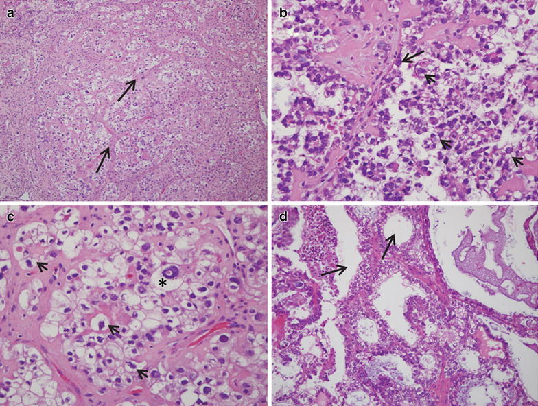
Background: We studied a primary culture developed from a biopsy of a clear cell carcinoma of the ovary (O-CCC) by (a) assessing its capacity to retain in vitro pathological features of the tumor of origin; (b) characterizing the main cells released from the complex mass without forced purification of any particular cellular entity; and (c) investigating its long-term proliferative capacity.

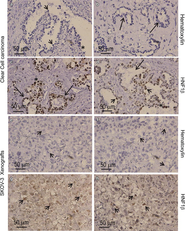
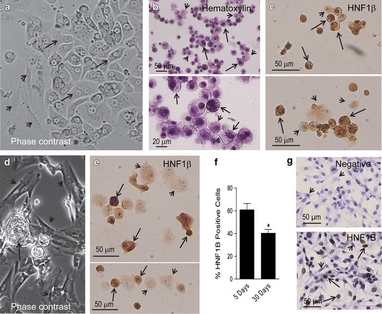
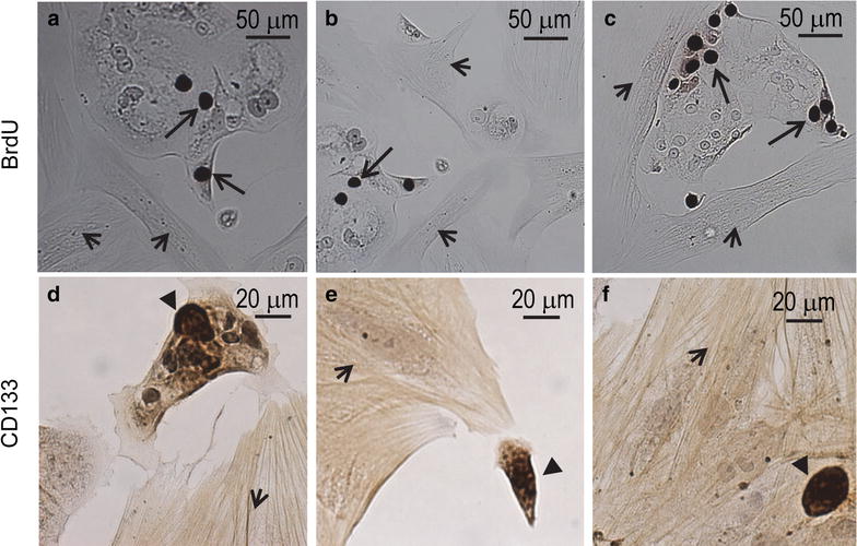
Methods: A primary cell culture was developed from a pelvic mass diagnosed as an O-CCC. The morphological analysis of the cell culture was carried out by phase contrast microscopy. Markers of epithelial, mesenchymal, and tumor initiating cells were evaluated by immunocytochemistry. Cell proliferation was studied by detection of bromodeoxyuridine (BrdU) incorporated into newly synthesized DNA. As a biomarker of O-CCC, we assessed the expression of hepatocyte nuclear factor (HNF) 1β.

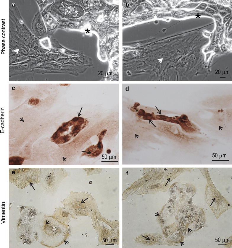
Results: We show that cells with epithelial morphological features express E-cadherin and expand with time in culture, a fact that the incorporation of BrdU confirms. Cells with mesenchymal-like characteristics that express the mesenchymal marker vimentin, however, allocate to the edges of the epithelial compartment. Moreover, we found that some cells with epithelial features also expressed vimentin. At the beginning of incubation, over 60 % of primary cells expressed the O-CCC marker HNF1β; such percentage declined upon passaging. We show that epithelial not mesenchymal cells undergo DNA replication, and that few cells in both epithelial and mesenchymal compartments express the stem-like tumor antigen CD133.

Conclusions: We provide proof-of-principle that cells separated in bulk from a biopsy of an O-CCC can be maintained in culture for several months, and that two consistent cellular compartments-one epithelial that retains the O-CCC marker HNF1β, and another mesenchymal-persist, and seem to have a cooperative interaction leading to the multiplication of epithelial cells within a mesenchymal cellular environment.

Keywords: E-cadherin; Epithelial; Hepatocyte nuclear factor 1β; Mesenchymal; Ovarian clear cell carcinoma; Vimentin.


Figures

Fig. 1
Fig. 1
Frozen sections of an O-CCC stained with H&E as diagnosed in the laboratory of pathology. a A field (original magnification ×10) containing abundant cells with clear cytoplasm, which are arranged around trabecular connective tissue (arrows). b (original magnification ×20) and c (original magnification ×40) denote the pathognomonic features of O-CCC, with hobnail cells (arrow in b), and large irregular cells with clear cytoplasm and eccentric prominent nuclei (arrowheads in b, c). The asterisk in c denotes a cell with clear cytoplasm, and excentric nucleus with a pseudo-inclusion. d An image of the tubulocystic aspect (arrows) of the clear carcinoma found in the omentum of the same patient
Fig. 2
Fig. 2
a, b Representative hematoxylin-stained, paraffin-embedded sections of the tumor biopsy utilized for isolating carcinoma cells for primary culture. Images depict a tubulopapillary pattern (arrows in b) with hobnail cells (arrowheads in a) and abundant cells with clear cytoplasm (asterisk in a). Positive immunohistochemical staining for HNF1β with hematoxylin counterstaining (c, d) in the biopsy are shown (arrows, tubuli with positive nuclei; arrowheads depicts positive nuclei in hobnail cells). Hematoxylin-stained sections (e, f), and HNF1β expression (g, h) in a xenograft of human SKOV-3 cells developed in immunosuppressed mice (arrowheads clear cells)
Fig. 3
Fig. 3
a Phase contrast image of a 5-day-old primary culture denoting a monolayer with an epithelial compartment depicting polygonal cells in a pavement-like arrangement (arrows) and a mesenchymal compartment (arrowheads) (original magnification ×20). b Cytospin cell preparations stained with hematoxylin and generated upon trypsinization of a section of a primary cell culture. The fields show two apparent types of cells of heterogeneous sizes, an abundant population of cells depicting dense chromatin and clear cytoplasm (arrows), and another population with less dense nuclei with prominent nucleolus, and less clear cytoplasm (arrowheads). c Cytospin cell preparations subjected to immunocytochemical staining for HNF1β. Many but not all cells show dark positive nuclei; arrows show positive-expressing cells whereas arrowheads show cells with negative expression; the panel also shows cells of different sizes. d Phase contrast image of a 30-day-old primary culture denoting higher proportion of cells with mesenchymal morphology (arrowheads)—when compared to a 5-day culture—always accompanied by epithelial-like cells growing in their vicinity (arrow) (original magnification ×10). e HNF1β staining in cytospin preparations of a 30-day-old primary culture displaying heterogeneity of expression, positive (arrows) or negative (arrowhead) cells. f Quantitation of the percentage of cells staining positive for HNF1β in cytospin preparations from 5-day or 30-day primary cultures of O-CCC. *p < 0.05 vs. 5 days (Student’s t-test). g Expression of HNF1β in a culture of SKOV-3 human cancer cells as counterstained with hematoxylin (upper panel negative; lower panel positive). Arrows depict positive immunostaining, whereas arrowheads indicate negative immunostaining
Fig. 4
Fig. 4
a, b Phase contrast images depicting the coexistence of epithelial (asterisks) and mesenchymal (arrowheads) cellular compartments after 8 weeks in culture. c, d E-cadherin immunoreactivity is shown in epithelial cells (arrows) but not in mesenchymal cells (arrowheads). e, f Vimentin immunostaining is positive mainly in the mesenchymal cells (arrows), yet some positivity is also observed in the cytoplasm of epithelial-like cells (arrowheads)
Fig. 5
Fig. 5
ac Bromodeoxyuridine (BrdU) was incorporated into cells of the epithelial compartment (arrows) but not into cells of the mesenchymal compartment (arrowheads). df CD133 positive cells (black triangles) associated to both the epithelial (d) and the mesenchymal (arrowheads) compartments (e, f)

References

    1. Domcke S, Sinha R, Levine DA, Sander C, Schultz N. Evaluating cell lines as tumour models by comparison of genomic profiles. Nat Commun. 2013;4:2126. doi: 10.1038/ncomms3126. - DOI - PMC - PubMed
    1. Lengyel E, Burdette JE, Kenny HA, Matei D, Pilrose J, Haluska P, Nephew KP, Hales DB, Stack MS. Epithelial ovarian cancer experimental models. Oncogene. 2014;33(28):3619–3633. doi: 10.1038/onc.2013.321. - DOI - PMC - PubMed
    1. Anglesio MS, Wiegand KC, Melnyk N, Chow C, Salamanca C, Prentice LM, Senz J, Yang W, Spillman MA, Cochrane DR, et al. Type-specific cell line models for type-specific ovarian cancer research. PLoS One. 2013;8(9):e72162. doi: 10.1371/journal.pone.0072162. - DOI - PMC - PubMed
    1. McGuire WP, Hoskins WJ, Brady MF, Kucera PR, Partridge EE, Look KY, Clarke-Pearson DL, Davidson M. Cyclophosphamide and cisplatin compared with paclitaxel and cisplatin in patients with stage III and stage IV ovarian cancer. N Engl J Med. 1996;334(1):1–6. doi: 10.1056/NEJM199601043340101. - DOI - PubMed
    1. Langdon SP, Lawrie SS. Establishment of ovarian cancer cell lines. Methods Mol Med. 2001;39:155–159. - PubMed

LinkOut - more resources