Cardiac involvement in Erdheim- Chester disease: MRI findings and literature revision
- PMID: 26405559
- PMCID: PMC4563377
- DOI: 10.1177/2058460115592273
Cardiac involvement in Erdheim- Chester disease: MRI findings and literature revision
Abstract
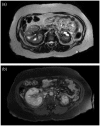
Erdheim-Chester disease (ECD) is a rare form of non-Langerhans cell histiocytosis, characterized by the involvement of several organs. The lesions may be skeletal or extra-skeletal: in particular, long bones, skin, lungs, and the cardiovascular and the central nervous systems can be affected. In this report, we describe a case of a 34-year-old man, who came to our observation with symptomatic ECD, for a correct assessment of the degree of cardiac involvement through magnetic resonance imaging (MRI).
Keywords: Erdheim-Chester disease; cardiac magnetic resonance; histiocytosis; non-Langerhans.
Figures


References
-
- Simiele N, Novoa F, Rodriguez N. Erdheim-Chester disease and Langerhans histiocytosis. A fortuitous association? Ann Med Int 2004; 21: 27–30. - PubMed
-
- Stoppacciaro A, Ferrarini M, Salmaggi C, et al. Immunohistochemical evidence of a cytokine and chemokine network in three patients with Erdheim-Chester disease. Implications for pathogenesis. Arthritis Rheum 2006; 54: 4018–4022. - PubMed
-
- Haroche J, Charlotte F, Arnaud L, et al. High prevalence of BRAF V600E mutations in Erdheim-Chester disease but not in other non-Langerhans cell histiocytoses. Blood 2012; 120: 2700–2703. - PubMed
Publication types
LinkOut - more resources
Full Text Sources
Other Literature Sources

