. 2015 Sep 24;163(1):84-94.
doi: 10.1016/j.cell.2015.08.055.

Sympathetic neuro-adipose connections mediate leptin-driven lipolysis

Affiliations

Sympathetic neuro-adipose connections mediate leptin-driven lipolysis

Wenwen Zeng et al. Cell. .

Abstract

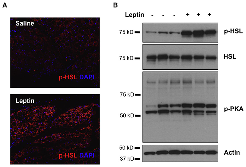
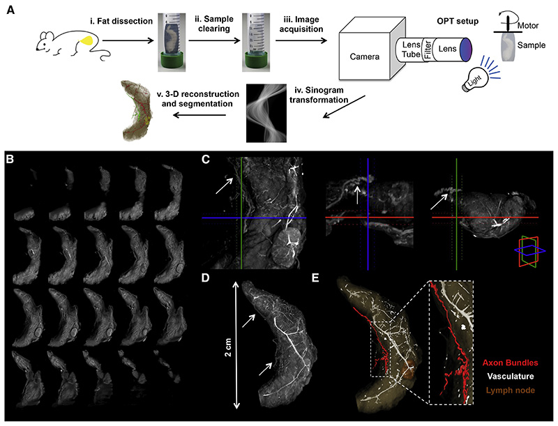
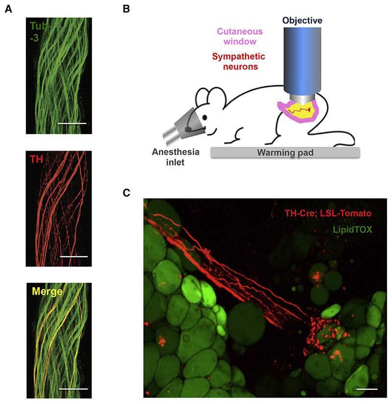
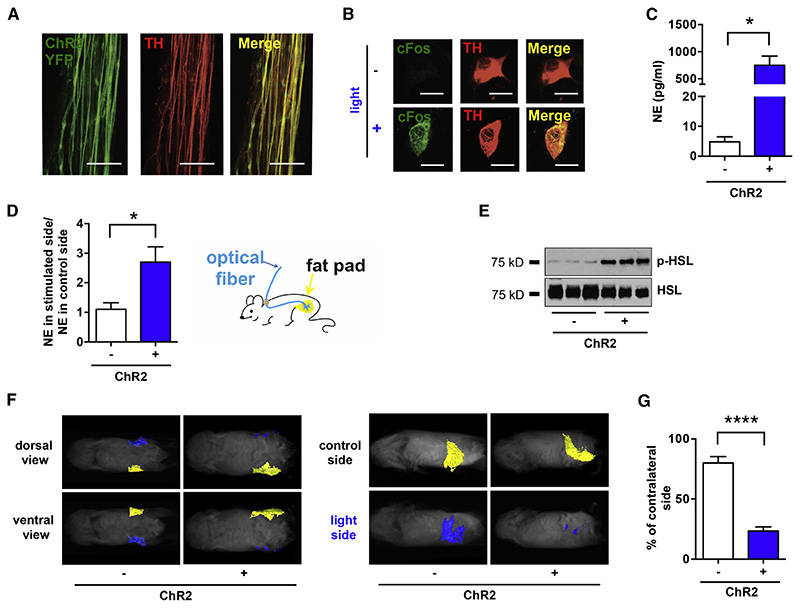
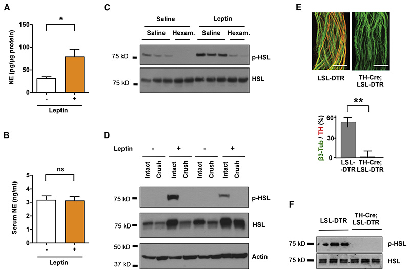
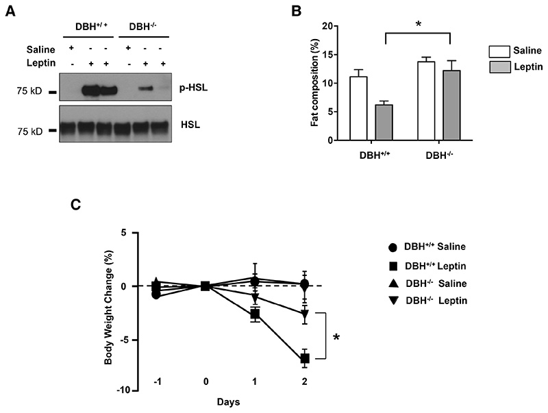
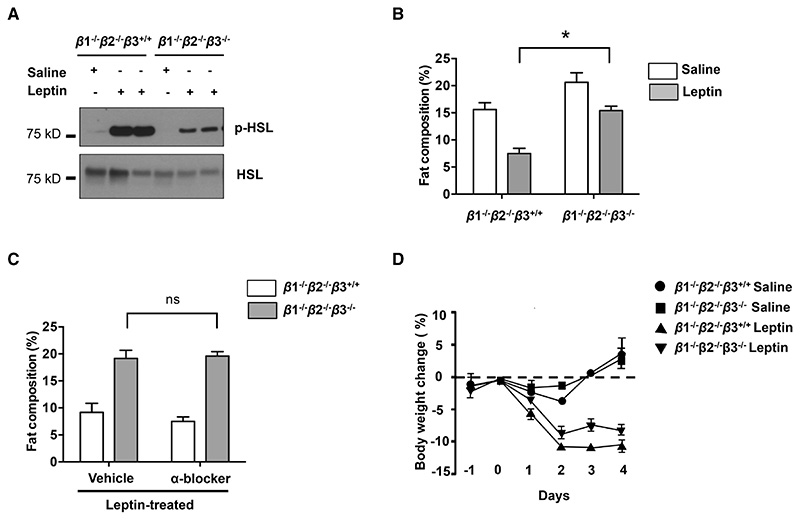
Leptin is a hormone produced by the adipose tissue that acts in the brain, stimulating white fat breakdown. We find that the lipolytic effect of leptin is mediated through the action of sympathetic nerve fibers that innervate the adipose tissue. Using intravital two-photon microscopy, we observe that sympathetic nerve fibers establish neuro-adipose junctions, directly "enveloping" adipocytes. Local optogenetic stimulation of sympathetic inputs induces a local lipolytic response and depletion of white adipose mass. Conversely, genetic ablation of sympathetic inputs onto fat pads blocks leptin-stimulated phosphorylation of hormone-sensitive lipase and consequent lipolysis, as do knockouts of dopamine β-hydroxylase, an enzyme required for catecholamine synthesis. Thus, neuro-adipose junctions are necessary and sufficient for the induction of lipolysis in white adipose tissue and are an efferent effector of leptin action. Direct activation of sympathetic inputs to adipose tissues may represent an alternative approach to induce fat loss, circumventing central leptin resistance. PAPERCLIP.


Figures

Figure 1
Figure 1. Leptin Stimulates HSL Phosphorylation in WAT.
(A) Immunostaining of phosphorylated HSL (red) in paraffin sections of epididymal fat of C57Bl6/J mice that were peripherally administrated with 500 ng/h recombinant leptin for 2 days. (B) Phosphorylated HSL and phosphorylated PKA substrates in total protein extracts of epididymal fats were examined by immunoblot analysis.
Figure 2
Figure 2. Neural Projections in Fat Detected with Optical Projection Tomography.
(A) Schematic representation of the OPT method applied to the subcutaneous inguinal fat. (i) Tissue dissection. (ii) Sample clearing. (iii) Image acquisition. (iv) Sinogram transformation. (v) 3-D reconstruction and segmentation. (B) OPT series of coronal sections of inguinal fat organ after 3-D reconstruction. (C) Orthogonal 400 μm OPT-slabs of inguinal fat in coronal, axial and sagittal view. Axon budles were identified based on the grey threshold level (arrows). (D) 3-D reconstruction in maximal intensity projection of the OPT coronal sections. (E) Surface view of segmented structures within inguinal fat.
Figure 3
Figure 3. Catecholaminergic Neurons Innervating Adipocytes Integrate Nerve Bundles of Mixed Molecular Identity.
(A) Partial co-localization of TH (red), an SNS marker, and Tub-3 (green), a general PNS marker, shown by immunohistochemistry of nerve bundles dissected from the inguinal fat pads of WT mice. Scale bar = 50 μm. (B) Schematic representation of the two-photon intra-vital imaging of neurons in the inguinal fat pad. (C) Intra-vital two-photon microscopy visualization of a neuro-adipose connection in the inguinal fat pad of a live TH-Cre; LSLS-Tomato mouse – LipidTOX (green) labels adipocytes. Scale bar = 100 μm.
Figure 4
Figure 4. Optogenetic Stimulation of SNS Neurons in Fat is Sufficient to Drive Lipolysis.
(A) Complete co-localization of YFP (green) and TH (red), shown by immunohistochemistry of nerve bundles dissected from the inguinal fat pads of TH-Cre; LSLS-ChR2-YFP. (B) C-Fos (green) induction in cultured SNS neurons after optogenetic activation. (C) Ex vivo NE release upon optogenetic stimulation of sympathetic SCG explants isolated from TH-Cre; LSL-ChR2-YFP mice and LSL-ChR2-YFP control mice (*p< 0.05, n=3-6). Results are shown as mean ± SEM. (D) In vivo NE release in subcutaneous fat upon optogenetic stimulation of sympathetic neurons in white adipose tissues of TH-Cre; LSL-ChR2-YFP and LSL-ChR2-YFP control mice that were subcutaneously implanted with optical fibers targeting the inguinal fat pad (*p<0.05, n=8). Results are shown as mean ± SEM. (E) Immunoblot analysis of phosphorylated HSL in total protein extracts of subcutaneous fats of TH-Cre; LSL-ChR2-YFP and LSL-ChR2-YFP control mice that were subcutaneously implanted with optical fibers targeting the inguinal fat pad and optogenetically stimulated for 2 weeks (details in Experimental Procedures section). (F) MRI-guided visualization of fat in TH-Cre; LSL-ChR2-YFP and LSL-ChR2-YFP control mice that were optogenetically stimulated for 4 weeks (yellow is control inguinal fat pat, blue is light-stimulated fat pad; details in Experimental Procedures section). (G) Quantification of fat reduction in stimulated side versus the contralateral control side (****p<0.0001, n=6). Results are shown as mean ± SEM. See also Movie S1 and S2.
Figure 5
Figure 5. Sympathetic Neurons are Locally Required for Leptin-induced Lipolysis.
C57BL6/J mice were peripherally administrated with 500 ng/h recombinant leptin or saline for 2 days. (A) NE content in subcutaneous fat pads (*p<0.05, n=5) and (B) NE serum levels were measured by NE ELISA (n=4). Results are shown as mean ± SEM. C57BL6/J mice were peripherally administrated with 500 ng/h recombinant leptin at 3 days after local crush injury of nerves in fat pads. (C) Phosphorylated HSL in total protein extracts of epididymal fats were examined by immunoblot analysis. (D) Fat pads in TH-Cre; LSL-DTR mice were locally treated with DT. Tissue specific ablation of SNS axons confirmed by immunostaining for Tub-3 and TH (**p< 0.001, n=6). Results are shown as mean ± SEM. (E) Immunoblot analysis of phosphorylated HSL in total protein extracts of subcutaneous fats of TH-Cre; LSL-DTR and control mice injected with DT, following leptin treatment (500 ng/h). See also Figure S1.
Figure 6
Figure 6. Norepinephrine Deficiency Impairs Leptin-induced Lipolysis.
(A) Immunoblot analysis of phosphorylated HSL in total protein extracts of fat pads of dopamine β-monoxygenase mutant and control littermates (DBH-/- and DBH+/+ respectively) mice that were treated with 500 ng/h recombinant leptin. (B) Whole-body fat composition (*p< 0.05). Results are shown as mean ± SEM (n=4-5). (C) Body weight change after leptin treatment (*p< 0.05). Results are shown as mean ± SEM (n=5).
Figure 7
Figure 7. Deficiency of all β-adrenergic Receptors Influences Leptin--induced Lipolysis.
(A) Immunoblot analysis of phosphorylated HSL in total protein extracts of fat pads of β1-/-β2-/-β3+/+ and β1-/-β2-/-β3-/- mice that were treated with 500 ng/h recombinant leptin. (B) Whole-body fat composition (*p< 0.05, n=4-5). Results are shown as mean ± SEM. (C) α-adrenergic receptors had a minor function in leptin-induced lipolysis (*p>0.05, n=4-5). Results are shown as mean ± SEM. (D) Whole-body fat composition of mice peripherally treated with recombinant leptin and α-blockers (phentolamine (5 mg/kg, IP) and phenoxybenzamine (10 mg/kg IP) was measured (n=4-5). Results are shown as mean ± SEM. See also Figure S2

Comment in

References

    1. Arvaniti K, Deshaies Y, Richard D. Effect of leptin on energy balance does not require the presence of intact adrenals. Am J Physiol. 1998;275:R105–111. - PubMed
    1. Awad S, Constantin-Teodosiu D, Macdonald IA, Lobo DN. Short-term starvation and mitochondrial dysfunction - a possible mechanism leading to postoperative insulin resistance. Clin Nutr. 2009;28:497–509. - PubMed
    1. Bachman ES, Dhillon H, Zhang CY, Cinti S, Bianco AC, Kobilka BK, Lowell BB. betaAR signaling required for diet-induced thermogenesis and obesity resistance. Science. 2002;297:843–845. - PubMed
    1. Bartness TJ, Kay Song C, Shi H, Bowers RR, Foster MT. Brain-adipose tissue cross talk. Proc Nutr Soc. 2005;64:53–64. - PubMed
    1. Bartness TJ, Song CK. Thematic review series: adipocyte biology. Sympathetic and sensory innervation of white adipose tissue. J Lipid Res. 2007;48:1655–1672. - PubMed

Publication types