Review
. 2015;5(4):699-713.
doi: 10.3233/JPD-150642.

The Synaptic Function of α-Synuclein

Free PMC article
Review

The Synaptic Function of α-Synuclein

Jacqueline Burré. J Parkinsons Dis. 2015.
Free PMC article

Abstract

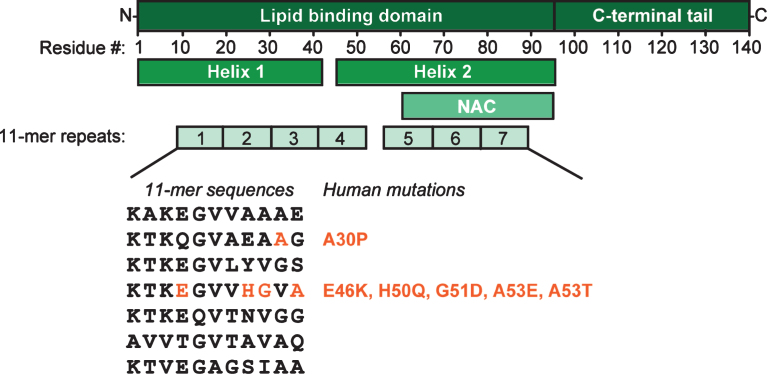
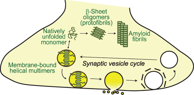
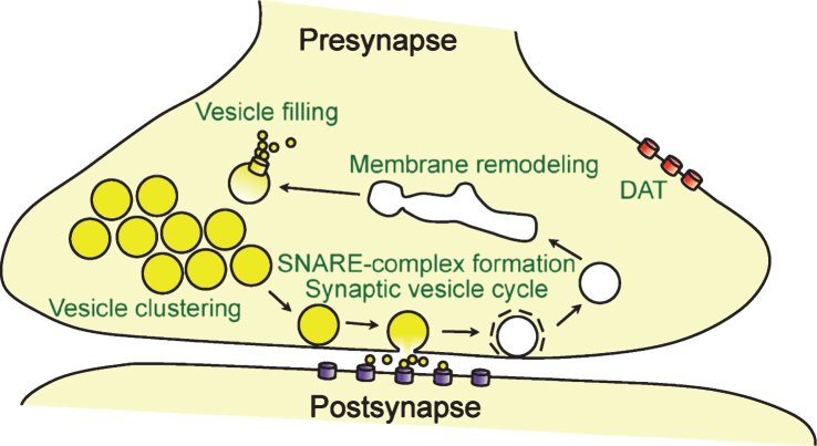
α-Synuclein is an abundant neuronal protein which localizes predominantly to presynaptic terminals, and is strongly linked genetically and pathologically to Parkinson's disease and other neurodegenerative diseases. While the accumulation of α-synuclein in the form of misfolded oligomers and large aggregates defines multiple neurodegenerative diseases called "synucleinopathies", its cellular function has remained largely unclear, and is the subject of intense investigation. In this review, I focus on the structural characteristics of α-synuclein, its cellular and subcellular localization, and discuss how this relates to its function in neurons, in particular at the neuronal synapse.

Keywords: SNARE; membranes; neurotransmitter release; synapse; synaptic vesicles; α-synuclein.


Figures

Fig.1
Fig.1
α-Synuclein domain structure. Upon binding to lipid membranes, the N-terminal domain of α-synuclein folds into two amphipathic helices; the C-terminal tail of α-synuclein does not contribute to membrane binding. The lipid binding domain can be divided into seven highly conserved 11-mer sequences. Helix 2 contains the aggregation-prone NAC-domain. All disease-linked mutations of α-synuclein are located in the second and fourth 11-mer stretch.
Fig.2
Fig.2
Physiological and pathological conformations of α-synuclein at the synapse. Cytosolic α-synuclein is monomeric and natively unfolded. Upon binding to synaptic vesicles, the N-terminal residues of α-synuclein adopt a helical structure. Membrane binding of α-synuclein is associated with its multimerization, which is essential for its physiological function at the synapse. Pathologically, unfolded α-synuclein in the cytosol can convert into β-sheet containing oligomers (protofibrils) which eventually form amyloid fibrils.
Fig.3
Fig.3
Function of α-synuclein at the synapse. Shown are the synaptic processes that α-synuclein has been reported to affect, including membrane remodeling, modulation of the dopamine transporter DAT and vesicular monoamine transporter VMAT2, clustering of synaptic vesicles and maintaining synaptic vesicle pools, promoting SNARE-complex assembly, and modulating the release cycle of synaptic vesicles.

References

    1. Maroteaux L, Campanelli JT, Scheller RH. Synuclein: A neuron-secific protein localized to the nucleus and presynaptic nerve terminal. J Neurosci. 1988;8:2804–2815. - PMC - PubMed
    1. Ueda K, Fukushima H, Masliah E, Xia Y, Iwai A, Yoshimoto M, Otero DA, Kondo J, Ihara Y, Saitoh T. Molecular cloning of cDNA encoding an unrecognized component of amyloid in Alzheimer disease. Proc Natl Acad Sci U S A. 1993;90:11282–11286. - PMC - PubMed
    1. Nakajo S, Omata K, Aiuchi T, Shibayama T, Okahashi I, Ochiai H, Nakai Y, Nakaya K, Nakamura Y. Purification and characterization of a novel brain-specific 14-kDa protein. J Neurochem. 1990;55:2031–2038. - PubMed
    1. Jakes R, Spillantini MG, Goedert M. Identification of two distinct synucleins from human brain. FEBS Lett. 1994;345:27–32. - PubMed
    1. Lavedan C, Leroy E, Dehejia A, Buchholtz S, Dutra A, Nussbaum RL, Polymeropoulos MH. Identification, localization and characterization of the human gamma-synuclein gene. Hum Genet. 1998;103:106–112. - PubMed

Publication types

Substances