. 2015 Sep 25;10(9):e0138835.
doi: 10.1371/journal.pone.0138835. eCollection 2015.

Expression of the Receptor Tyrosine Kinase EphB2 on Dendritic Cells Is Modulated by Toll-Like Receptor Ligation but Is Not Required for T Cell Activation

Affiliations

Expression of the Receptor Tyrosine Kinase EphB2 on Dendritic Cells Is Modulated by Toll-Like Receptor Ligation but Is Not Required for T Cell Activation

Patrice N Mimche et al. PLoS One. .

Abstract

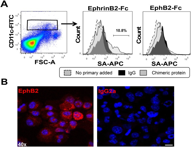
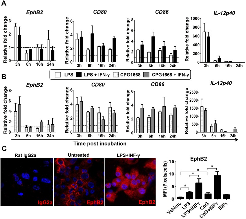
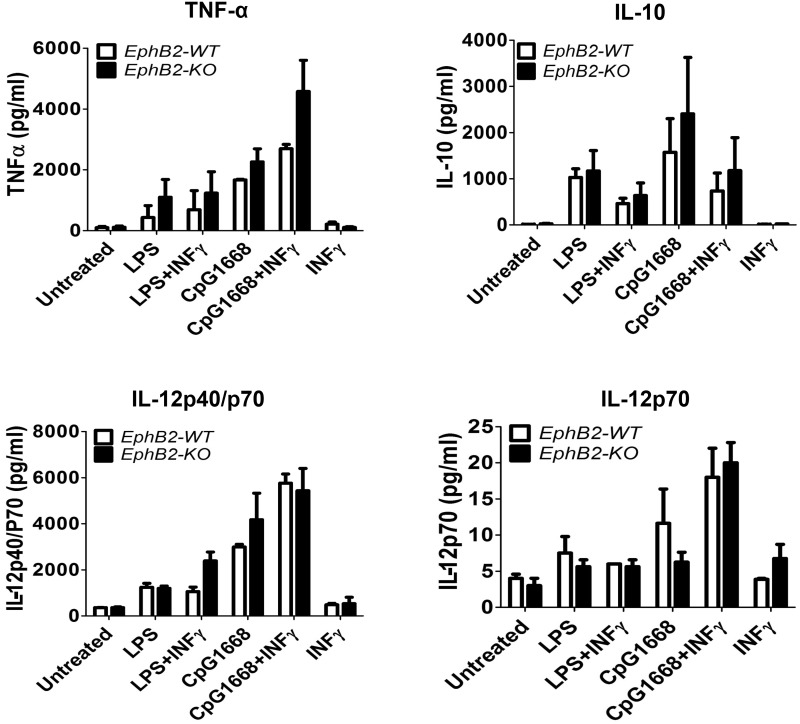
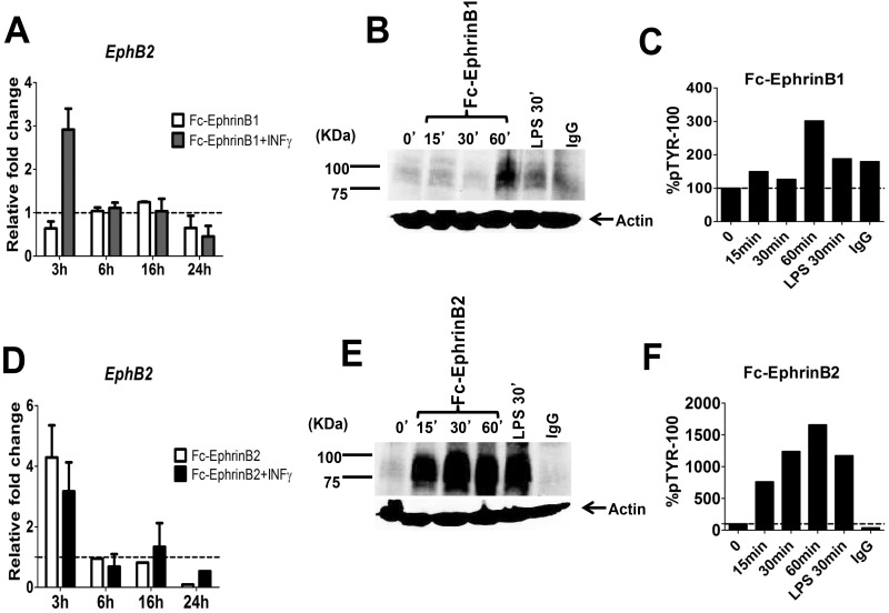
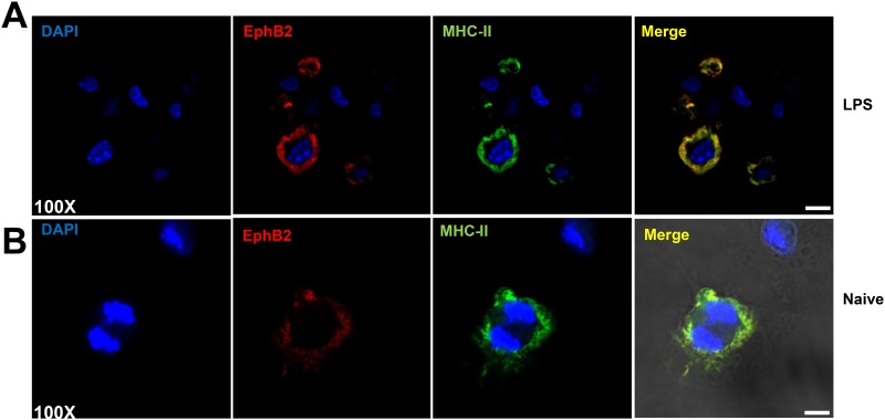
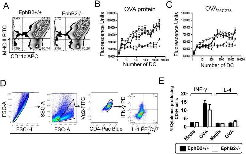
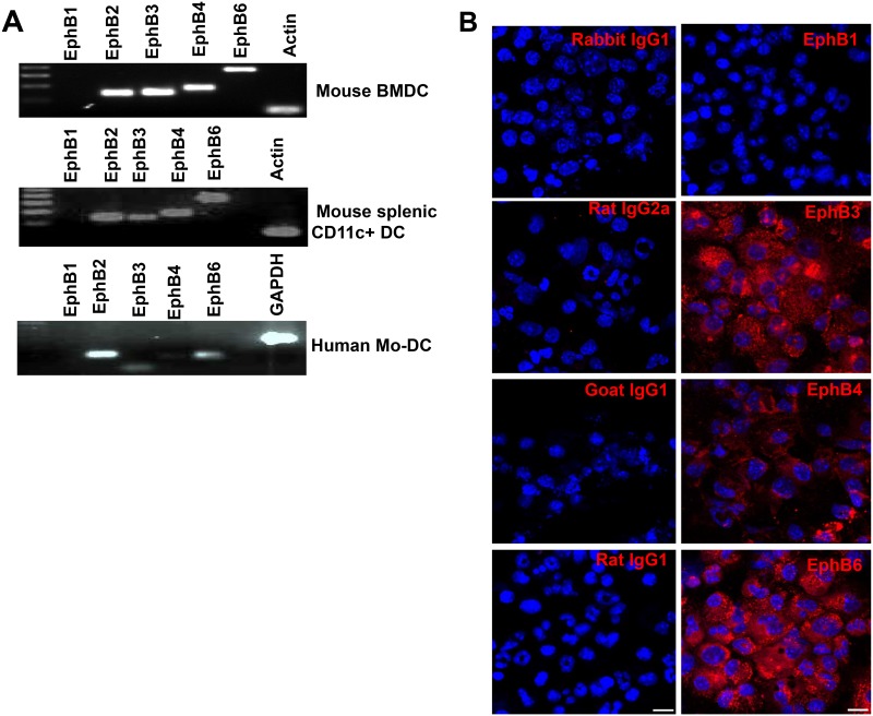
The Eph receptor tyrosine kinases interact with their ephrin ligands on adjacent cells to facilitate contact-dependent cell communication. Ephrin B ligands are expressed on T cells and have been suggested to act as co-stimulatory molecules during T cell activation. There are no detailed reports of the expression and modulation of EphB receptors on dendritic cells, the main antigen presenting cells that interact with T cells. Here we show that mouse splenic dendritic cells (DC) and bone-marrow derived DCs (BMDC) express EphB2, a member of the EphB family. EphB2 expression is modulated by ligation of TLR4 and TLR9 and also by interaction with ephrin B ligands. Co-localization of EphB2 with MHC-II is also consistent with a potential role in T cell activation. However, BMDCs derived from EphB2 deficient mice were able to present antigen in the context of MHC-II and produce T cell activating cytokines to the same extent as intact DCs. Collectively our data suggest that EphB2 may contribute to DC responses, but that EphB2 is not required for T cell activation. This result may have arisen because DCs express other members of the EphB receptor family, EphB3, EphB4 and EphB6, all of which can interact with ephrin B ligands, or because EphB2 may be playing a role in another aspect of DC biology such as migration.


Conflict of interest statement

Competing Interests: The authors have declared that no competing interests exist.

Figures

Fig 1
Fig 1. Splenic CD11chi and bone marrow-derived DCs express EphB2.
(A) Eph receptor expression on the surface of naïve splenic CD11chi DCs was detected by incubation with Ephrin-B2-Fc chimeric protein and binding was detected using a biotinylated anti-Fc antibody and streptavidin-APC (SA-APC) using flow cytometry. (B) The expression of EphB2 on BMDCs incubated with anti-EphB2 antibody or isotype control (IgG2a) detected by immunofluorescence. Magnification 40x; Scale bar, 10μm.
Fig 2
Fig 2. EphB2 expression on BMDCs can be modulated by ligation with Toll-like receptor ligands.
(A) BMDC were incubated with a Toll Like receptor (TLR)-4 agonist (lipopolysaccharide (LPS) 1μg/ml) and (B) a TLR-9 agonist (CpG1668 1μM) with or without recombinant mouse interferon (IFN)-γ at a concentration of 20ng/ml. EphB2 mRNA was quantified by qPCR at different time points post-stimulation. The modulation of transcription for known co-stimulatory molecules CD80 and CD86 as well as the cytokine interleukin (IL)-12p40 in response to TLR stimulation in the same experiments are shown for comparison. (C) The change in EphB2 protein expression at 22 hours post-incubation with LPS and IFN-γ is shown and the mean fluorescence quantified for different conditions. All graphs represent the median value of pooled data across 3 independent BMDC preparations ±SD and data analyzed using One-way ANOVA– Kruskal Wallis test and Dunn’s multiple comparisons post-test. *P<0.05. MFI = Mean Fluorescence Intensity.
Fig 3
Fig 3. Stimulation of BMDCs from EphB2+/+ and EphB2-/- mice with TLR receptor agonists results in similar levels of secreted cytokine.
BMDCs from EphB2+/+ and EphB2-/- mice were incubated with TLR ligands LPS and CpG1668 with and without the addition of recombinant interferon (IFN)-γ for 20 hours. Interleukin (IL)-12p40 (A), IL-12p70 (B), IL-10 (C) and tumor necrosis factor (TNF)-α (D) were measured in the culture supernatant using Luminex. The graphs are representative of 3 individual experiments and bars represent the mean ±SD of 2 replicate wells plated.
Fig 4
Fig 4. BMDCs stimulated with EphrinB2 upregulate EphB2 mRNA.
Naïve BMDCs were plated onto plate-bound Ephrin-B1-Fc fusion protein (A, B, C) or plate bound Ephrin-B2-Fc fusion protein (D, E, F). The transcription of EphB2 with and without the addition of recombinant interferon-γ (IFN-γ) was monitored by qPCR over 24 hours of culture (A, D). The efficiency of the Eph receptor ligation was monitored by assessing tyrosine phosphorylation on DCs by western blot (B, E) and the phosphorylation quantified using densitometry (C, F). All graphs represent the median value of pooled data from 3 independent DC preparations ±SD.
Fig 5
Fig 5. EphB2 co-localizes with MHC-II on BMDCs.
Two examples are shown: (A) Representative BMDCs stimulated with 1μg/ml lipopolysaccharide (LPS) and 20ng/ml recombinant mouse interferon (IFN)-γ for 22 hours; (B) shows unstimulated cells. EphB2 is shown in red (Northernlights557), MHC-II is shown in green (FITC) and DAPI is shown to demarcate the nuclei of the cells. Magnification 100x; Scale bar, 20μm.
Fig 6
Fig 6. EphB2 is not required for development of Th1 cells in culture.
BMDCs were matured from mouse bone marrow (A) and incubated with the MF2.2D9 T cell hybridoma and chicken ovalbumin (OVA) (B) or OVA257-278 peptide (C) for 18 hours. The amount of IL-2 secreted by the MF2.2D9 T cell hybridoma was measured using HT-2 cells and the cell number enumerated using an ATP-luminescence assay. The graphs represent the mean value ±SD of 3 replicate wells for each dilution of EphB2-/- BMDCs (gray lines) or littermate control BMDCs (black lines). Circles represent no addition of a stimulus and stimulated BMDCs are represented by squares. (D) BMDCs derived from bone marrow of EphB2-/- or EphB2+/+ mice were incubated with splenic CD4+ T cells purified from naïve OT-II T cell receptor transgenic mice (n = 5) and OVA protein for 5 days. The development and expansion of Th1 and Th2 cells were measured by intracellular staining of interferon (IFN)-γ and interleukin (IL)-4 respectively (E). All graphs are representative of at least 2 experiments, with the exception of (E) which is the median value of pooled data ±SD from 3 separate preparations of BMDCs.
Fig 7
Fig 7. DCs express Eph receptors other than EphB2.
RNA was extracted from naïve bone marrow-derived DCs (A), splenic DCs (B) and human monocyte-derived DCs (C) and PCR performed using primers specific for mouse and human EphB receptors. The protein expression of the EphB receptor family was detected using immunohistochemistry on naïve mouse BMDCs (D) and the expression compared to that detected using isotype controls. Magnification 40x; Scale bar, 10μm.

References

    1. Hirai H, Maru Y, Hagiwara K, Nishida J, Takaku F. A novel putative tyrosine kinase receptor encoded by the eph gene. Science. 238(4834):1717–1720. . - PubMed
    1. Aasheim H-C, Delabie J, Finne EF. Ephrin-A1 binding to CD4+ T lymphocytes stimulates migration and induces tyrosine phosphorylation of PYK2. Blood. 2005; 105(7):2869–2876. . - PubMed
    1. de Saint-Vis B, Bouchet C, Gautier G, Valladeau J, Caux C, Garrone P. Human dendritic cells express neuronal Eph receptor tyrosine kinases: role of EphA2 in regulating adhesion to fibronectin. Blood. 2003; 102(13):4431–4440. - PubMed
    1. Holen HL, Nustad K, Aasheim H-C. Activation of EphA receptors on CD4+CD45RO+ memory cells stimulates migration. Journal of Leukocyte Biology. 2010; 87(6):1059–68. 10.1189/jlb.0709497 . - DOI - PubMed
    1. Jellinghaus S, Poitz DM, Ende G, Augstein A, Weinert S, Stütz B, et al. Ephrin-A1/EphA4-mediated adhesion of monocytes to endothelial cells. Biochimica et Biophysica Acta (BBA)—Molecular Cell Research. 2013; 2013 October;1833(10):2201–11. 10.1016/j.bbamcr.2013.05.017 . - DOI - PubMed

Publication types

MeSH terms