. 2015 Sep 25;11(9):e1005179.
doi: 10.1371/journal.ppat.1005179. eCollection 2015 Sep.

DYRK2 Negatively Regulates Type I Interferon Induction by Promoting TBK1 Degradation via Ser527 Phosphorylation

Affiliations

DYRK2 Negatively Regulates Type I Interferon Induction by Promoting TBK1 Degradation via Ser527 Phosphorylation

Tai An et al. PLoS Pathog. .

Abstract

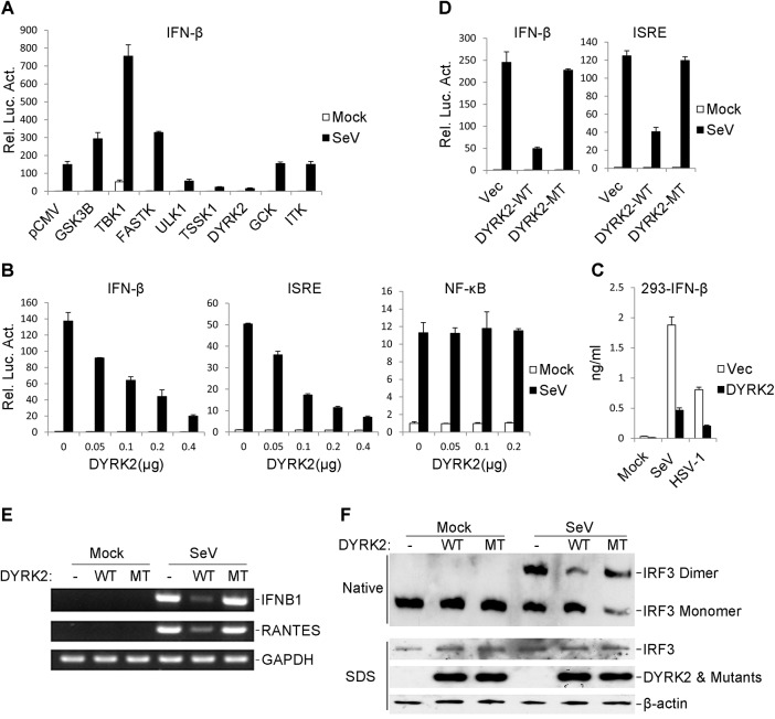
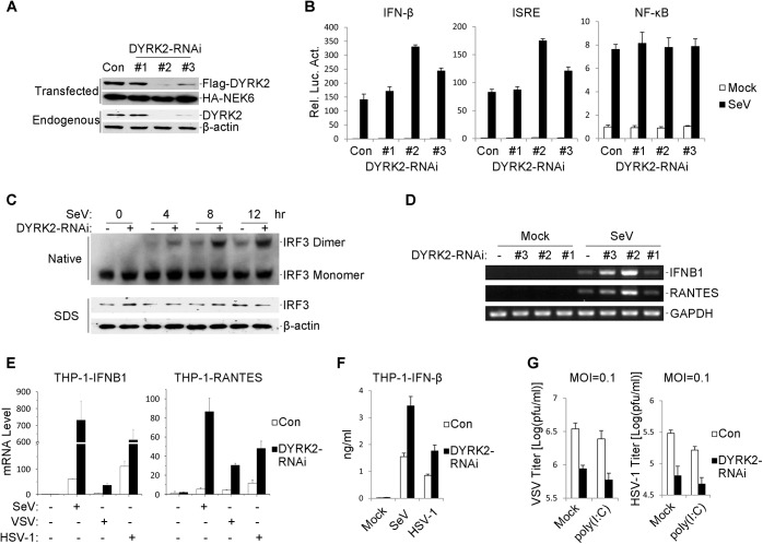
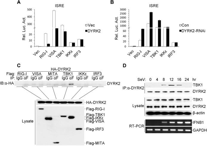
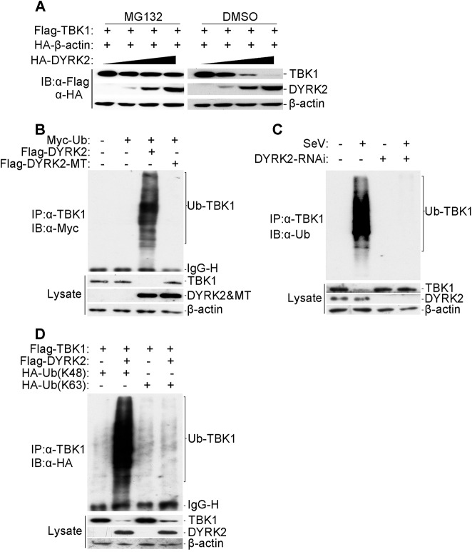
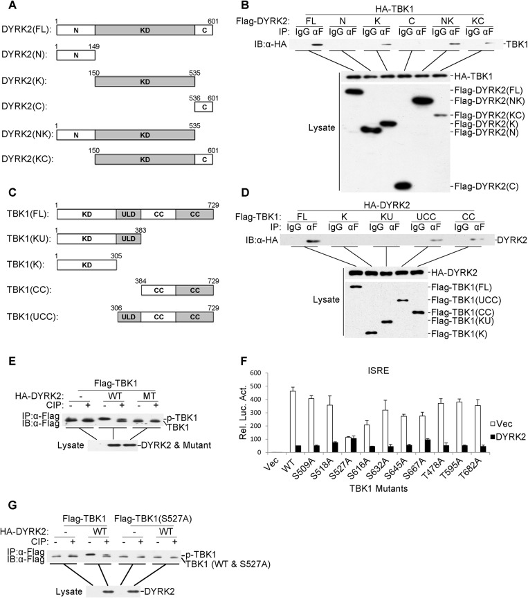
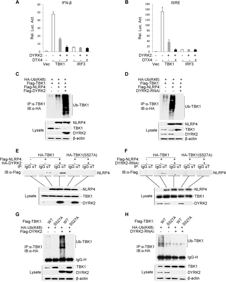
Viral infection activates the transcription factors NF-κB and IRF3, which contribute to the induction of type I interferons (IFNs) and cellular antiviral responses. Protein kinases play a critical role in various signaling pathways by phosphorylating their substrates. Here, we identified dual-specificity tyrosine-(Y)-phosphorylation-regulated kinase 2 (DYRK2) as a negative regulator of virus-triggered type I IFN induction. DYRK2 inhibited the virus-triggered induction of type I IFNs and promoted the K48-linked ubiquitination and degradation of TANK-binding kinase 1 (TBK1) in a kinase-activity-dependent manner. We further found that DYRK2 phosphorylated Ser527 of TBK1, which is essential for the recruitment of NLRP4 and for the E3 ubiquitin ligase DTX4 to degrade TBK1. These findings suggest that DYRK2 negatively regulates virus-triggered signaling by targeting TBK1 for phosphorylation and priming it for degradation, and these data provide new insights into the molecular mechanisms that dictate the cellular antiviral response.


Conflict of interest statement

The authors have declared that no competing interests exist.

Figures

Fig 1
Fig 1. The overexpression of DYRK2 markedly inhibited the virus-triggered activations of IRF3 and IFNB1 gene transcription.
(A) Identification of DYRK2 as an inhibitor of SeV-induced IFN-β activation. The 293 cells were individually transfected with human cDNA clones (Origene, Inc.) and the IFN-β luciferase reporter. Twenty hours after transfection, the cells were treated with or without SeV for 10 h before the luciferase assays were performed. (B) DYRK2 inhibited the SeV-induced activations of the ISRE and the IFN-β promoter but not NF-κB in dose-dependent manners in the 293 cells. The 293 cells (1×10⁵) were transfected with the ISRE, NF-κB reporter or IFN-β promoter luciferase plasmids (0.1 μg) and the indicated amount of DYRK2 expression plasmid. Twenty hours after transfection, the cells were infected with or without SeV for 10 h before the luciferase assays were performed. The graphical data are presented as the means ± the SDs (n = 3). (C) Effects of DYRK2 on the SeV- and HSV-1-induced secretion of IFN-β. The 293 cells (1×10⁵) were transfected with the indicated plasmids. After 20 h of incubation, the cells were infected with SeV or HSV-1 for 12 h. The culture medium was collected for quantification of the indicated cytokines by ELISA. (D) Wild-type DYRK2 but not mutant DYRK2 suppressed the SeV-induced activations of the ISRE and the IFN-β promoter. The 293 cells (1×10⁵) were transfected with the ISRE or IFN-β promoter luciferase plasmids (0.1 μg) and an expression plasmid for the wild-type DYRK2 (DYRK2-WT) or the kinase-dead DYRK2 mutant (DYRK2-MT) (0.1 μg). Twenty hours after transfection, the cells were or were not infected with SeV for 10 h before the luciferase assays were performed. The graphical data are presented as the means ± the SDs (n = 3). (E) DYRK2 inhibited the SeV-induced endogenous gene transcription of IFNB1 and RANTES. The 293 cells (2×10⁵) were transfected with the indicated plasmids (0.2 μg each) for 20 h and then infected or not infected with SeV for 10 h before reverse transcription PCR was performed. (F) DYRK2 inhibited the SeV-induced dimerization of IRF3. The 293 cells (2×10⁵) were transfected with the indicated plasmids. Twenty hours after transfection, the cells were infected with or without SeV for 10 h. Cell lysates were separated by native (top) or SDS (bottom) PAGE and analyzed by immunoblotting with the indicated antibodies.
Fig 2
Fig 2. Effects of RNAi-mediated knockdown of DYRK2 on SeV-induced signaling and IRF3 activation.
(A) Effects of DYRK2 RNAi on the expression of transfected and endogenous DYRK2. In the upper panel, 293 cells (2×10⁵) were transfected with expression plasmids for Flag-DYRK2 and HA-NEK6 (0.1 μg each) and the indicated RNAi plasmids (1 μg). Twenty-four hours after transfection, the cell lysates were analyzed by immunoblotting with the indicated antibodies. In the lower panel, the 293 cells (2×10⁵) were transfected with the control or indicated DYRK2 RNAi plasmids (1 μg each) for 36 h. The cell lysates were analyzed by immunoblotting with the indicated antibodies. (B) Effects of DYRK2 RNAi on the SeV-induced activations of the ISRE, NF-κB and the IFN-β promoter. The 293 cells (1×10⁵) were transfected with the ISRE, NF-κB or IFN-β promoter reporters (0.05 μg) and the indicated RNAi plasmids (0.5 μg each) for 36 h and then infected or not infected with SeV for 10 h before the luciferase assays were performed. The graphical data are presented as the means ± the SDs (n = 3). (C) Knockdown of DYRK2 promoted SeV-induced IRF3 dimerization. The 293 cells (2×10⁵) were transfected with control or DYRK2 RNAi (#2) plasmids (1 μg). Thirty-six hours after transfection, the cells were infected with or without SeV for 0, 4, 8, or 12 h. The cell lysates were separated by native (top) or SDS (bottom) PAGE, and the blots were analyzed using the indicated antibodies. (D) Effects of DYRK2 RNAi on the SeV-induced endogenous gene transcriptions of IFNB1 and RANTES. The 293 cells (2×10⁵) were transfected with the indicated RNAi plasmids (1 μg each) for 36 h and then infected or not infected with SeV for 10 h before reverse transcription PCR was performed. (E) Effects of DYRK2 RNAi on SeV-, VSV- and HSV-1-induced transcriptions of IFNB1 and RANTES in THP-1 cells. RNAi-transduced stable THP-1 cells (2×10⁵) were infected or not infected with SeV/VSV/HSV-1 for 8 h before qPCR was performed. (F) Effects of DYRK2 RNAi on SeV- and HSV-1-induced secretion of IFN-β. RNAi-transduced stable THP-1 cells (1×10⁵) were infected with SeV or HSV-1 for 12 h. The culture medium was collected for quantitation of the indicated cytokines by ELISA. (G) Effects of DYRK2 RNAi on virus replication. RNAi-transduced stable THP-1 cells (1×10⁵) were mock-transfected or transfected with poly(I:C) (1μg) for 16 h and then infected with VSV or HSV-1 (MOI = 0.1). The supernatants were harvested 24 h after infection for standard plaque assays.
Fig 3
Fig 3. DYRK2-mediated virus-triggered signaling at the level of TBK1.
(A) The overexpression of DYRK2 inhibited RIGI-, VISA-, and TBK1-mediated signaling but not IKKε- and IRF3-mediated signaling. The 293 cells (1×10⁵) were transfected with DYRK2 or control plasmids (0.1 μg) together with ISRE luciferase and the indicated plasmids (0.1 μg each). Luciferase assays were performed 20 h after transfection. The graphical data are presented as the means ± the SDs (n = 3). (B) Knockdown of DYRK2 promoted ISRE activation by RIGI, VISA, and TBK1 but not by IKKε or IRF3. The 293 cells (1×10⁵) were transfected with control or DYRK2 RNAi (#2) plasmids (0.5 μg). After 20 h, the cells were selected with puromycin (1 μg/ml) for 24 h and then re-transfected with the ISRE luciferase and the indicated expression plasmids (0.1 μg each). Reporter assays were performed 24 h after transfection. The graphical data are presented as the means ± the SDs (n = 3.) (C) DYRK2 interacted with TBK1 but not with RIG-I, VISA, MITA, IKKε, or IRF3 in the overexpression system. The 293 cells (2×10⁶) were transfected with the indicated plasmids (5 μg each). Coimmunoprecipitation and immunoblotting analyses were performed with the indicated antibodies (upper). The expressions of the transfected proteins were analyzed by immunoblotting with anti-HA or anti-Flag antibodies (lower). (D) Kinetics of the DYRK2–TBK1 association after viral infection. The 293 cells (1×10⁸) were treated with MG132 and infected with SeV for the indicated times. Small fractions of the cells were prepared for RT-PCR (lower). The remaining cell fractions were lysed, and the lysates were coimmunoprecipitated with an anti-DYRK2 antibody or preimmune serum. The coimmunoprecipitates were analyzed by immunoblotting with anti-TBK1 or anti-DYRK2 antibodies (upper). The expression levels of endogenous TBK1, DYRK2, and β-actin were detected by immunoblot analyses with the indicated antibodies (middle).
Fig 4
Fig 4. DYRK2 promoted TBK1 degradation via K48-linked ubiquitination.
(A) Overexpression of DYRK2 induced TBK1 degradation in a dose-dependent manner. The 293T cells (2×10⁵) were transfected with the Flag-TBK1, HA-β-actin and HA-DYRK2 plasmids (0.1, 0.2 or 0.4 μg) and were treated with dimethyl sulfoxide (DMSO) or MG132. The cells were lysed, and the lysates were analyzed by immunoblotting with anti-Flag or anti-HA antibodies. (B) Overexpression of wild-type DYRK2 but not mutant DYRK2, promoted the ubiquitination of TBK1. The 293 cells (1×10⁷) were transfected with the indicated plasmids. Twenty-four hours after transfection, cell lysates were immunoprecipitated with an anti-TBK1 antibody. The immunoprecipitates were analyzed by immunoblotting with an anti-Myc antibody (upper). Protein expression was analyzed by immunoblotting with the indicated antibodies (lower). (C) Effects of DYRK2 RNAi on the SeV-induced ubiquitination of endogenous TBK1. The 293 cells (5×10⁷) were transfected with control or DYRK2 RNAi (#2) plasmids. Twenty hours after transfection, the cells were infected or not infected with SeV for 10 h. The cell lysates were immunoprecipitated with an anti-TBK1 antibody. The immunoprecipitates were analyzed by immunoblotting with an anti-ubiquitin antibody (top). The expressions of related proteins were examined by immunoblotting with the indicated antibodies (bottom). (D) DYRK2 promoted K48-linked but not K63-linked ubiquitination of TBK1. The 293 cells (2×10⁶) were transfected with HA-tagged Lys-48-only or Lys-63-only ubiquitin plasmids and the other indicated plasmids. Twenty-four hours after transfection, cell lysates were immunoprecipitated with an anti-TBK1 antibody and then analyzed by immunoblotting with an anti-HA antibody (upper panel). The expressions of related proteins were examined by immunoblotting with the indicated antibodies (lower panel).
Fig 5
Fig 5. DYRK2 phosphorylated TBK1 at S527.
(A and C) Schematics of human DYRK2 (A) and TBK1 (C) and their truncation mutants. (B) DYRK2 interacted with TBK1 via its kinase domain. The 293 cells (2×10⁶) were transfected with the indicated plasmids (5 μg each). Coimmunoprecipitations were performed with anti-Flag or IgG (control). The immunoprecipitates were analyzed by immunoblotting with an anti-HA antibody (top). The expressions of the transfected proteins were analyzed by immunoblotting with anti-HA (middle) or anti-Flag (bottom) antibodies. (D) DYRK2 bound to the coiled-coil domain of TBK1. The 293 cells (2×10⁶) were transfected with the indicated plasmids and then treated and analyzed as described in (B). (E) Wild-type DYRK2 but not mutant DYRK2 promoted TBK1 phosphorylation. The 293 cells (2×10⁶) were transfected with the indicated plasmids (5 μg each). Cell lysates were immunoprecipitated with an anti-Flag antibody, treated with or without calf intestine phosphatase (CIP), and analyzed by immunoblotting with an anti-Flag antibody (top). The expressions of the transfected proteins were analyzed by immunoblotting with an anti-HA antibody (bottom). (F) The effects of DYRK2 on the wild-type and mutant TBK1 as determined by ISRE activation. The 293 cells (1×10⁵) were transfected with an ISRE reporter plasmid (0.1 μg) and the indicated expression plasmids (0.1 μg each) for 20 h before the luciferase assays were performed. The graphical data are presented as the means ± the SDs (n = 3). (G) DYRK2 promoted the phosphorylation of wild-type TBK1 but not the TBK1 S527A mutant. The 293 cells (2×10⁶) were transfected with the indicated plasmids and then treated and analyzed as described in (E).
Fig 6
Fig 6. Effects of DYRK2 on the NLRP4-mediated inhibition of Type I interferon induction.
(A and B) The synergistic inhibition of DTX4 on the activation of the IFN-β promoter (A) and ISRE (B). The 293 cells (1×10⁵) were transfected with the indicated plasmids. Luciferase assays were performed 20 h after transfection. The graphical data are presented as the means ± the SDs (n = 3). (C and D) DYRK2 overexpression potentiated (C) whereas DYRK2 knockdown inhibited (D) the degradation of TBK1 that was induced by NLRP4. The 293 cells (2×10⁶) were transfected with the indicated plasmids. Twenty-four hours after transfection, cell lysates were immunoprecipitated with an anti-TBK1 antibody. The immunoprecipitates were analyzed by immunoblotting with an anti-HA antibody (upper). Protein expression was analyzed by immunoblotting with the indicated antibodies (lower). (E and F) DYRK2 overexpression enhanced (E) whereas DYRK2 knockdown abolished (F) the interaction between TBK1 and NLRP4. The 293 cells (2×10⁶) were transfected with the indicated plasmids and then treated with (E) or without (F) MG132. Cell lysates were analyzed with the indicated antibodies (upper). The expression of the transfected proteins was analyzed by immunoblotting with anti-HA and anti-Flag antibodies (lower). (G and H) DYRK2 promoted the degradation of wild-type TBK1 but not of the S527A mutant. The 293 cells (2×10⁶) were transfected with the indicated plasmids and then treated and analyzed as described in (C and D).

References

    1. Akira S, Uematsu S, Takeuchi O (2006) Pathogen recognition and innate immunity. Cell 124: 783–801. - PubMed
    1. Kumar H, Kawai T, Akira S (2011) Pathogen recognition by the innate immune system. Int Rev Immunol 30: 16–34. 10.3109/08830185.2010.529976 - DOI - PubMed
    1. O'Neill LA (2008) The interleukin-1 receptor/Toll-like receptor superfamily: 10 years of progress. Immunol Rev 226: 10–18. 10.1111/j.1600-065X.2008.00701.x - DOI - PubMed
    1. Takeuchi O, Akira S (2010) Pattern recognition receptors and inflammation. Cell 140: 805–820. 10.1016/j.cell.2010.01.022 - DOI - PubMed
    1. Hoebe K, Du X, Georgel P, Janssen E, Tabeta K, et al. (2003) Identification of Lps2 as a key transducer of MyD88-independent TIR signalling. Nature 424: 743–748. - PubMed

Publication types

MeSH terms