. 2015 Sep 25;10(9):e0138870.
doi: 10.1371/journal.pone.0138870. eCollection 2015.

Targeting Rapamycin to Podocytes Using a Vascular Cell Adhesion Molecule-1 (VCAM-1)-Harnessed SAINT-Based Lipid Carrier System

Affiliations

Targeting Rapamycin to Podocytes Using a Vascular Cell Adhesion Molecule-1 (VCAM-1)-Harnessed SAINT-Based Lipid Carrier System

Ganesh Ram R Visweswaran et al. PLoS One. .

Abstract

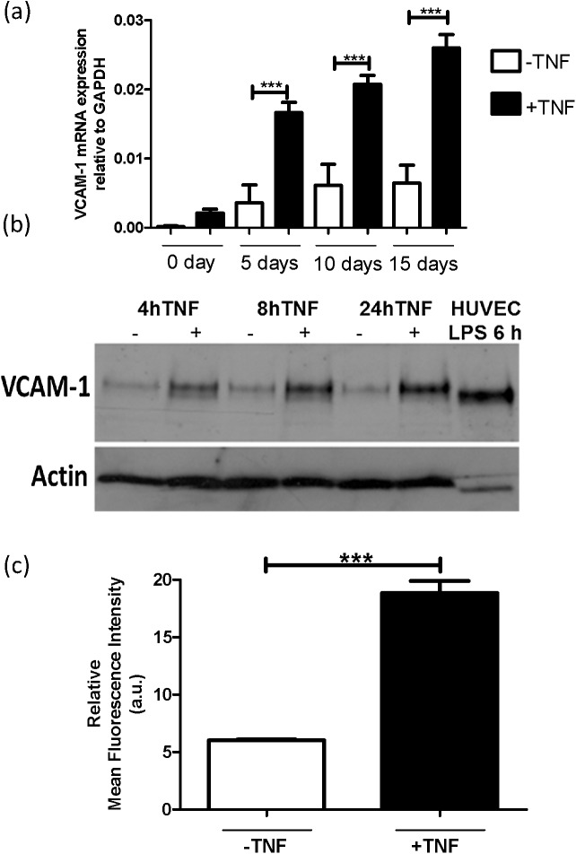
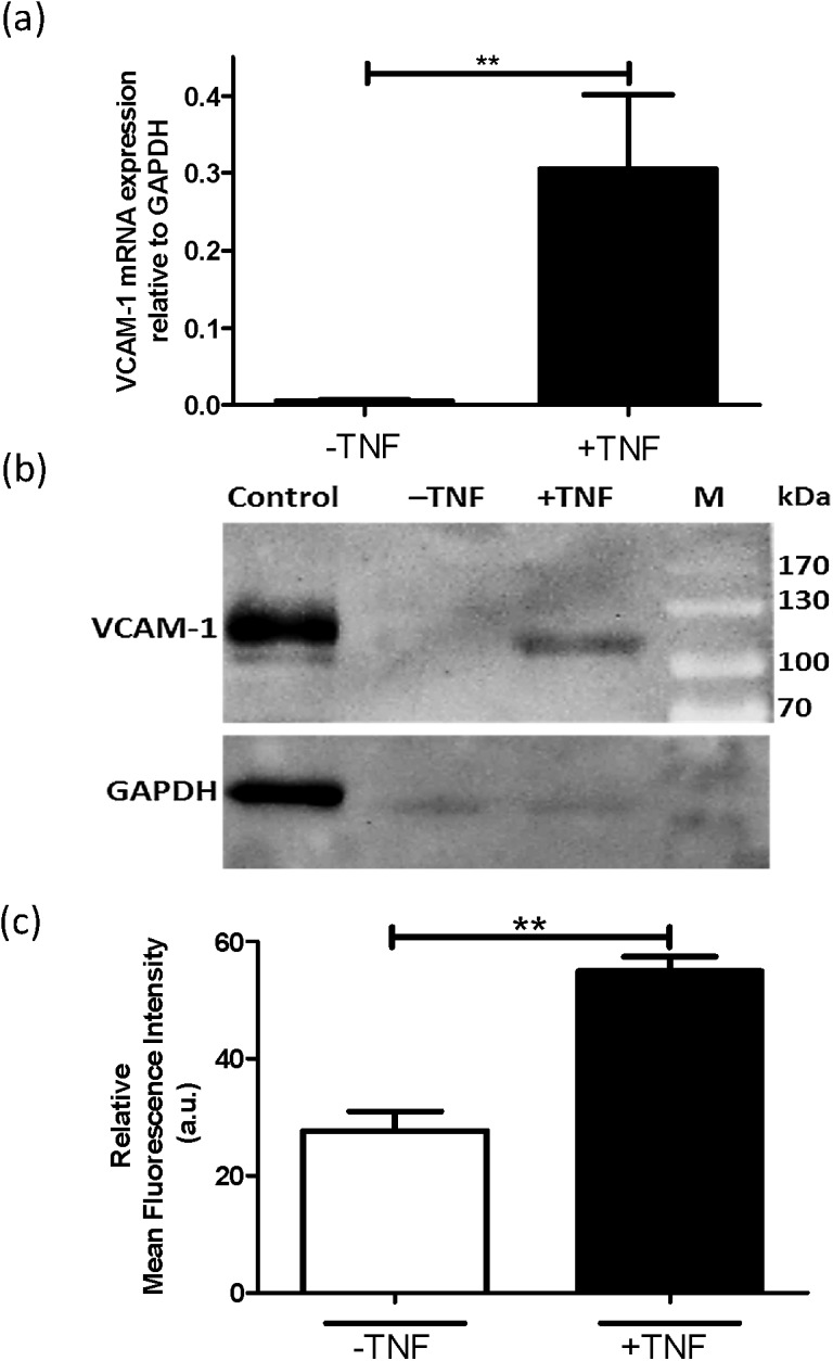
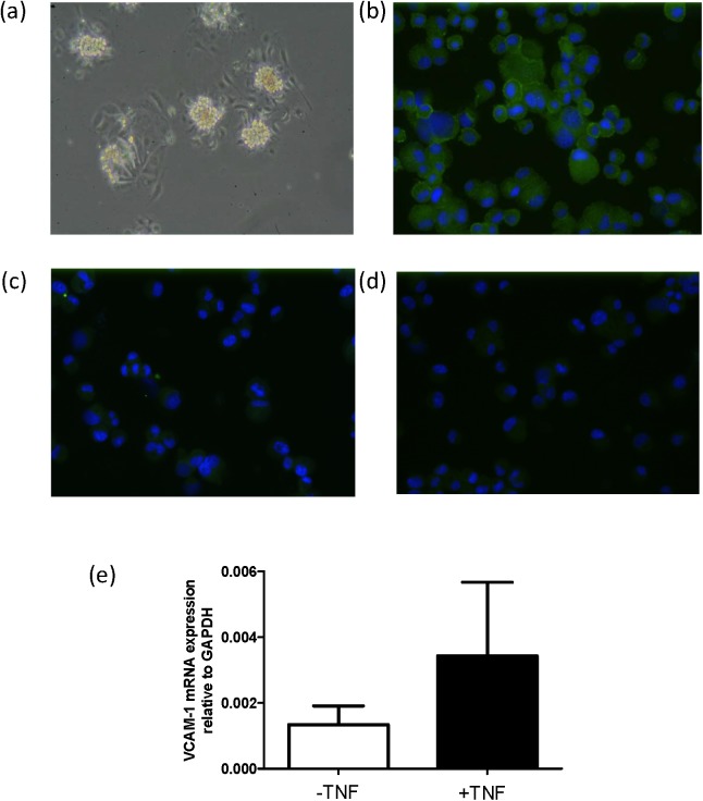
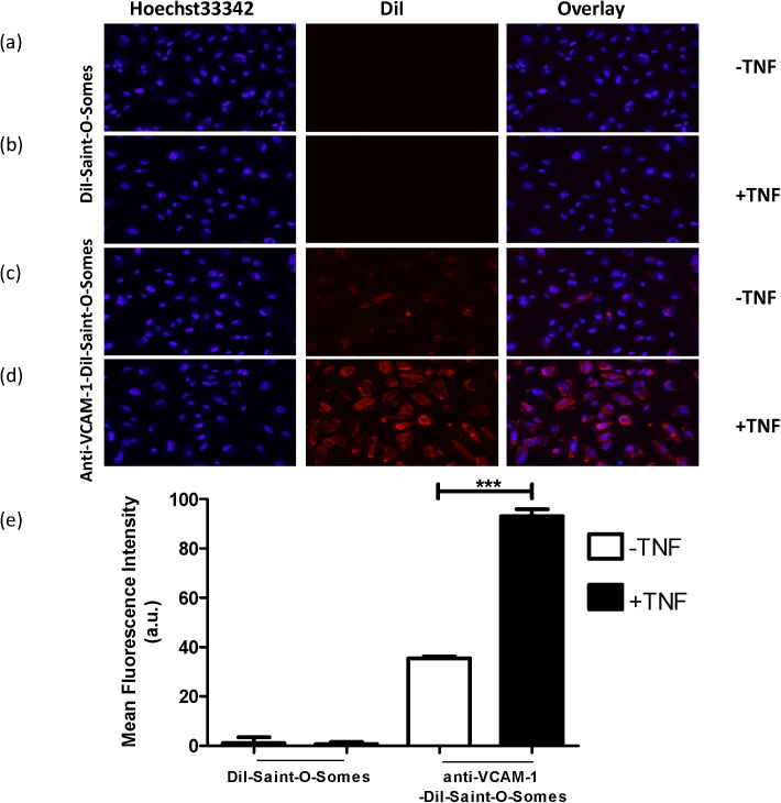
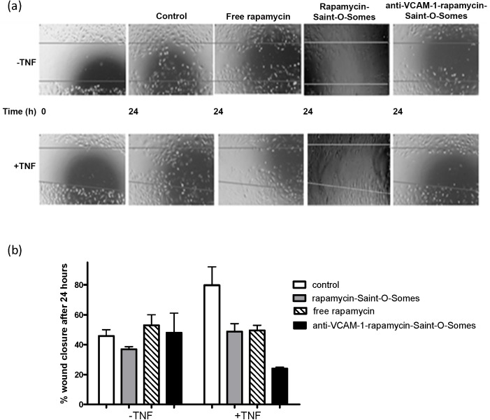
Together with mesangial cells, glomerular endothelial cells and the basement membrane, podocytes constitute the glomerular filtration barrier (GFB) of the kidney. Podocytes play a pivotal role in the progression of various kidney-related diseases such as glomerular sclerosis and glomerulonephritis that finally lead to chronic end-stage renal disease. During podocytopathies, the slit-diaphragm connecting the adjacent podocytes are detached leading to severe loss of proteins in the urine. The pathophysiology of podocytopathies makes podocytes a potential and challenging target for nanomedicine development, though there is a lack of known molecular targets for cell selective drug delivery. To identify VCAM-1 as a cell-surface receptor that is suitable for binding and internalization of nanomedicine carrier systems by podocytes, we investigated its expression in the immortalized podocyte cell lines AB8/13 and MPC-5, and in primary podocytes. Gene and protein expression analyses revealed that VCAM-1 expression is increased by podocytes upon TNFα-activation for up to 24 h. This was paralleled by anti-VCAM-1 antibody binding to the TNFα-activated cells, which can be employed as a ligand to facilitate the uptake of nanocarriers under inflammatory conditions. Hence, we next explored the possibilities of using VCAM-1 as a cell-surface receptor to deliver the potent immunosuppressant rapamycin to TNFα-activated podocytes using the lipid-based nanocarrier system Saint-O-Somes. Anti-VCAM-1-rapamycin-SAINT-O-Somes more effectively inhibited the cell migration of AB8/13 cells than free rapamycin and non-targeted rapamycin-SAINT-O-Somes indicating the potential of VCAM-1 targeted drug delivery to podocytes.


Conflict of interest statement

Competing Interests: The authors have declared that no competing interests exist.

Figures

Fig 1
Fig 1. VCAM-1 is expressed in AB8/13 cells and the expression is increased upon TNFα-activation.
(a) RT-qPCR analysis of human VCAM-1 mRNA expression. The AB8/13 cells were differentiated for 0, 5, 10, and 15 days at 37°C and treated with TNFα (10 ng/ml) for 4 h. (b) Western blot analysis of VCAM-1 (~110-kDa) and actin (~42-kDa) (loading control) proteins expression in cell lysate of AB8/13 cells, 15 days differentiated at 37°C in the absence (-) or presence (+) of TNFα for various time points. (c) FACS analysis of VCAM-1 protein in TNFα-activated (10 ng/ml; 24 h) and quiescent AB8/13 cells incubated with mouse anti-human-VCAM-1 for 45 min at 4°C followed by incubation with fluorescently-labelled secondary antibody. *p <0.001.
Fig 2
Fig 2. VCAM-1 is expressed by the mouse podocyte cell line MPC-5.
(a) mRNA expression of VCAM-1 in the absence (-) or presence (+) of TNFα (10 ng/ml; 24 h) as analyzed by RT-qPCR analysis. (b) Western blot analysis of protein extracts of MPC-5 cells that were incubated in the absence (-) or presence (+) of TNFα (10 ng/ml; 24 h) prior to harvesting. M and control indicate respectively, pre-stained molecular weight marker and positive control (HUVEC activated with LPS for 6 h). (c) FACS analysis of VCAM-1 expression by MPC-5 cells using anti-VCAM-1 antibody by flow cytometry. *p <0.001.
Fig 3
Fig 3. VCAM-1 is expressed in primary mouse podocytes.
(a) Phase-contrast view of glomerular outgrowths of isolated glomeruli from mouse kidney, 2 days after seeding in podocyte-specific medium. Immuno-staining of cytospots of the ICAM-2 negative fraction after ICAM-2 re-beading, detecting nephrin (b), while the endothelial marker Tie2 (c) is not detected. Panel (d) shows background staining with control rabbit-IgG antibodies (and does not detect anything). Antibodies that specifically bound to the cells are stained in green. Nuclei were stained in blue with DAPI. (e) VCAM-1 mRNA expression in mouse primary podocytes in the absence (-) or presence (+) of TNFα, as determined by RT-qPCR. Data are presented as mean values +/- sd, n = 3 from three independent experiments.
Fig 4
Fig 4. Anti-VCAM-1-DiI-Saint-O-Somes bind to AB8/13 cells.
DiI-SAINT-O-Somes (a, b) and anti-VCAM-1-DiI-SAINT-O-Somes (c, d) were incubated for 4 h at 37°C with AB8/13 cells in the absence (-) (A, C) or presence (+) (b, d) of TNFα (10 ng/ml; 24 h). The nuclei were stained blue with Hoechst 33342. Original magnification 200x. (e) Quantification of binding of anti-VCAM-1-DiI-SAINT-O-Somes or DiI-SAINT-O-Somes to AB8/13 cells. Cells incubated in equal volumes of HN buffer were taken as controls and their values were subtracted from test samples. The mean fluorescence intensity (MFI) was determined by FACS and analyzed by FlowJoTM. *p <0.001.
Fig 5
Fig 5. Effect of anti-VCAM-1-rapamycin-SAINT-O-Somes on AB8/13 cells.
(a) A scratch was made into confluent cell cultures of AB8/13 in the absence (-) or presence (+) of TNFα (10 ng/ml; 24 h). These cells were first treated with HN buffer (control), free drug, rapamycin-SAINT-O-Somes and anti-VCAM-1-rapamycin-SAINT-O-Somes for 12 h, after which a wound was induced and the wound closure was followed in time up to 24 h. (b) The number of cells that had migrated into the scratch area (═) were quantified from two independent experiments by ImageJ.

References

    1. Leeuwis JW, Nguyen TQ, Dendooven A, Kok RJ, Goldschmeding R (2010) Targeting podocyte-associated diseases. Adv Drug Deliv Rev 62: 1325–1336. 10.1016/j.addr.2010.08.012 - DOI - PubMed
    1. Siddiqi FS, Advani A (2013) Endothelial-podocyte crosstalk: the missing link between endothelial dysfunction and albuminuria in diabetes. Diabetes 62: 3647–3655. 10.2337/db13-0795 - DOI - PMC - PubMed
    1. Haraldsson B, Jeansson M (2009) Glomerular filtration barrier. Curr Opin Nephrol Hypertens 18: 331–335. 10.1097/MNH.0b013e32832c9dba - DOI - PubMed
    1. Chiang WC, Geel TM, Altintas MM, Sever S, Ruiters MH, Reiser J (2010) Establishment of protein delivery systems targeting podocytes. PLoS ONE 5: e11837 10.1371/journal.pone.0011837 - DOI - PMC - PubMed
    1. Hauser PV, Pippin JW, Kaiser C, Krofft RD, Brinkkoetter PT, Hudkins KL, et al. (2010) Novel siRNA delivery system to target podocytes in vivo. PLoS ONE 5: e9463 10.1371/journal.pone.0009463 - DOI - PMC - PubMed

Publication types

LinkOut - more resources