. 2015 Dec;81(24):8315-29.
doi: 10.1128/AEM.02587-15. Epub 2015 Sep 25.

Bioinformatic characterization of glycyl radical enzyme-associated bacterial microcompartments

Affiliations

Bioinformatic characterization of glycyl radical enzyme-associated bacterial microcompartments

Jan Zarzycki et al. Appl Environ Microbiol. 2015 Dec.

Abstract

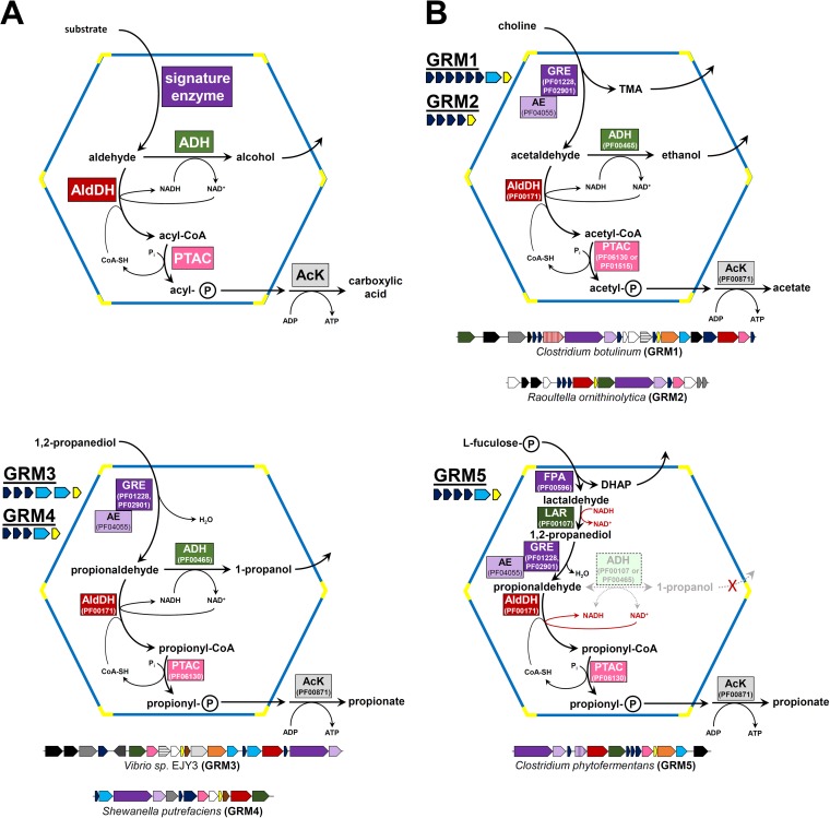
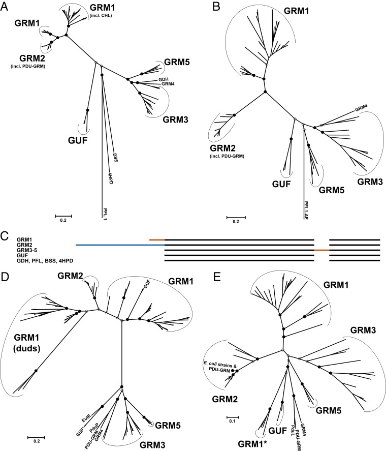
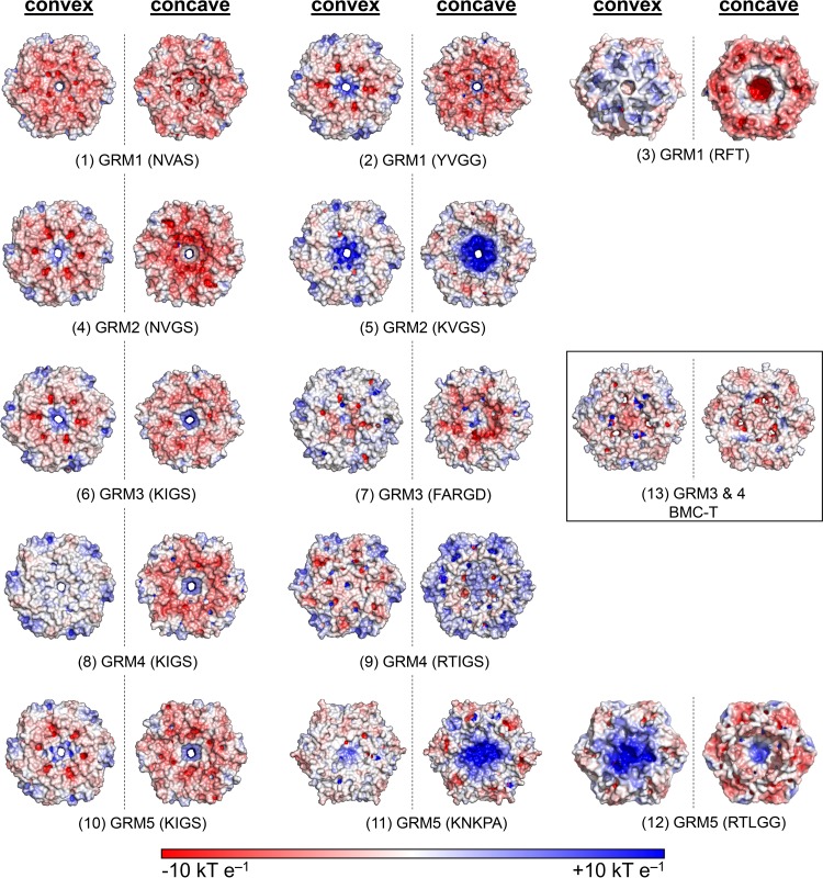
Bacterial microcompartments (BMCs) are proteinaceous organelles encapsulating enzymes that catalyze sequential reactions of metabolic pathways. BMCs are phylogenetically widespread; however, only a few BMCs have been experimentally characterized. Among them are the carboxysomes and the propanediol- and ethanolamine-utilizing microcompartments, which play diverse metabolic and ecological roles. The substrate of a BMC is defined by its signature enzyme. In catabolic BMCs, this enzyme typically generates an aldehyde. Recently, it was shown that the most prevalent signature enzymes encoded by BMC loci are glycyl radical enzymes, yet little is known about the function of these BMCs. Here we characterize the glycyl radical enzyme-associated microcompartment (GRM) loci using a combination of bioinformatic analyses and active-site and structural modeling to show that the GRMs comprise five subtypes. We predict distinct functions for the GRMs, including the degradation of choline, propanediol, and fuculose phosphate. This is the first family of BMCs for which identification of the signature enzyme is insufficient for predicting function. The distinct GRM functions are also reflected in differences in shell composition and apparently different assembly pathways. The GRMs are the counterparts of the vitamin B12-dependent propanediol- and ethanolamine-utilizing BMCs, which are frequently associated with virulence. This study provides a comprehensive foundation for experimental investigations of the diverse roles of GRMs. Understanding this plasticity of function within a single BMC family, including characterization of differences in permeability and assembly, can inform approaches to BMC bioengineering and the design of therapeutics.


Figures

FIG 1
FIG 1
Schematics of the functions of metabolosomes. (A) Generalized functional scheme for metabolism by experimentally characterized catabolic bacterial microcompartments. The signature enzyme generates a toxic and/or volatile aldehyde, which is successively metabolized by an acylating aldehyde dehydrogenase (AldDH), a phosphotransacylase (PTAC), and an acyl kinase (AcK). The cosubstrates CoA and NAD+ are regenerated by the PTAC and an alcohol dehydrogenase (ADH) within the lumen of the compartment. (B) Schematics of the proposed functions of the different GRE-associated microcompartments (GRMs). The GREs in GRM1 and GRM2 are predicted to be choline TMA-lyases or ethanolamine ammonia-lyases, whereas the GREs in GRM3 to GRM5 are proposed to function as 1,2-propanediol dehydratases. GRM5 BMC loci also encode a fuculose phosphate aldolase (FPA) and a putative lactaldehyde reductase (LAR). The function of the lactaldehyde reductase would negate the necessity for a propanol dehydrogenase. Representative gene clusters for each type of locus are depicted below the schematic with the corresponding coloring: purple, signature enzymes; purple hatched, FPA; red, AldDH; red hatched, inactive (dud) AldDH; green, ADH; pink, PTAC; light gray, AcK; orange, PduS (PF13375, PF13534, PF01512, PF10531); gray hatched, EutJ (PF11104); brown, DUF336 (PF03928); dark gray, putative transporters (various Pfams); black, putative regulators (various Pfams); white, other ancillary genes. The average number and types of BMC shell proteins of the different GRMs are also indicated; dark blue, BMC-H; light blue, BMC-T; yellow, BMC-P.
FIG 2
FIG 2
Phylogenetic trees of GRM-associated enzymes. Maximum likelihood trees are drawn to scale, and branch lengths are based on the number of substitutions per site. Bootstrap values for important nodes are represented as filled circles (above 50%) and empty circles (below 50%). (A) GRE tree generated from 83 amino acid sequences. The sequences used for calibration were those of biochemically characterized GREs not associated with BMCs: vitamin B12-independent glycerol dehydratase (GDH) from Clostridium butyricum, the benzylsuccinate synthase (BSS) from Thauera aromatica, the 4-hydroxyphenylacetate decarboxylase (4HPD) from Clostridium difficile, and pyruvate formate-lyase type 1 (PFL1) from E. coli. PDU-GRM, the locus in Escherichia fergusonii which appears to be a fusion of the PDU BMC and GRM loci; GUF, GREs of unknown function; CHL, choline TMA-lyase. (B) Tree of the activating enzymes inferred from 79 amino acid sequences. The activating enzyme of the pyruvate formate-lyase type 1 from E. coli (PFL1-AE) was used as an outgroup. (C) Cartoon representation of the multiple-sequence alignment of the GREs. Orange segments, extensions/insertions that likely constitute EPs with linkers; blue segment, the N-terminal extension of the GREs of the GRM2 BMCs that appears to be a partial domain duplication. (D) Tree of the acylating aldehyde dehydrogenases (AldDHs) inferred from 111 amino acid sequences. The AldDH homologs from the conventional ethanolamine-utilizing BMC (EutE) and propanediol-utilizing BMC (PduP) from Salmonella enterica were included in the analysis. GRM1 (duds), presumably inactive AldDH homologs; GUF*, AldDHs from D. psychrophila. (E) Tree of PduL-like phosphotransacylase homologs inferred from 74 amino acid sequences. The phosphotransacylase (PduL) from the experimentally characterized PDU BMC of S. enterica was included for comparison. GRM1*, the PduL homologs of Desulfovibrio spp.
FIG 3
FIG 3
Electrostatic surface renderings of homology models of shell protein oligomers conserved among GRM loci. Both the concave and convex sides of the model of each shell protein assembly are shown. The GRM type (pore motifs) are indicated. The boxed GRM3 BMC-T serves as an example for shell protein trimers with three pores. Positive surface potentials (blue) and negative surface potentials (red) ranging from 10 to −10 kT/e are indicated, with the Boltzmann's constant (k), temperature (T), and the charge of an electron (e).

References

    1. Lower BH, Bazylinski DA. 2013. The bacterial magnetosome: a unique prokaryotic organelle. J Mol Microbiol Biotechnol 23:63–80. doi:10.1159/000346543. - DOI - PubMed
    1. Gorby YA, Beveridge TJ, Blakemore RP. 1988. Characterization of the bacterial magnetosome membrane. J Bacteriol 170:834–841. - PMC - PubMed
    1. Kerfeld CA, Heinhorst S, Cannon GC. 2010. Bacterial microcompartments. Annu Rev Microbiol 64:391–408. doi:10.1146/annurev.micro.112408.134211. - DOI - PubMed
    1. Chowdhury C, Sinha S, Chun S, Yeates TO, Bobik TA. 2014. Diverse bacterial microcompartment organelles. Microbiol Mol Biol Rev 78:438–468. doi:10.1128/MMBR.00009-14. - DOI - PMC - PubMed
    1. Axen SD, Erbilgin O, Kerfeld CA. 2014. A taxonomy of bacterial microcompartment loci constructed by a novel scoring method. PLoS Comput Biol 10:e1003898. doi:10.1371/journal.pcbi.1003898. - DOI - PMC - PubMed

Publication types

MeSH terms

LinkOut - more resources