

Surgical History of Sleep Apnea in Pediatric Patients with Chiari Type 1 Malformation
- PMID: 26408064
- PMCID: PMC4584091
- DOI: 10.1016/j.nec.2015.06.009
Surgical History of Sleep Apnea in Pediatric Patients with Chiari Type 1 Malformation
Abstract
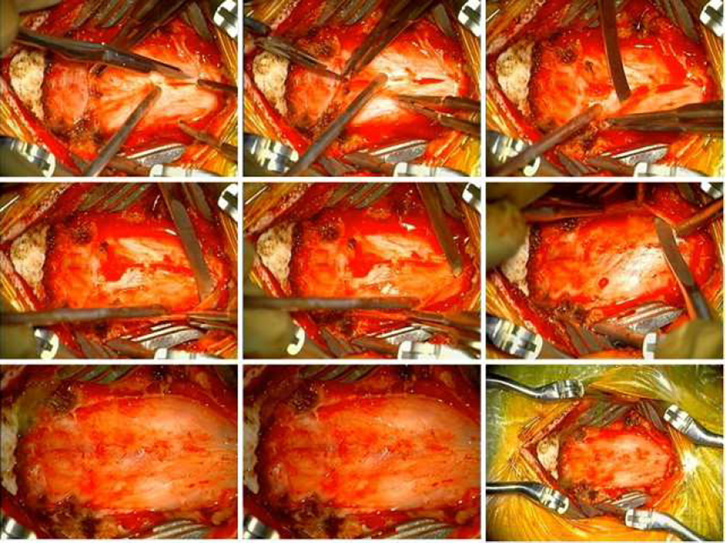
Sleep apnea represents a relative indication for posterior fossa decompression in pediatric patients with Chiari malformation type 1. Duraplasty was associated with improvement of sleep apnea in 100% of patients and dural splitting with improvement in 50% of patients. Duraplasty and dural splitting were associated with a similar reduction in tonsillar herniation on radiographic imaging of 58% (37% excluding tonsillectomy) and 35%, respectively. Longitudinal follow-up studies of patients with either neurologic deficits or severe symptoms will further elucidate the natural history of Chiari malformation type 1 and more appropriately gauge the risk-benefit tradeoff of surgical intervention.
Keywords: Chiari malformation; Dural splitting; Duraplasty; Sleep apnea.
Copyright © 2015 Elsevier Inc. All rights reserved.
Figures


Similar articles
-
Natural and surgical history of Chiari malformation Type I in the pediatric population.J Neurosurg Pediatr. 2016 Mar;17(3):343-52. doi: 10.3171/2015.7.PEDS1594. Epub 2015 Nov 20. J Neurosurg Pediatr. 2016. PMID: 26588459
-
Posterior fossa decompression without duraplasty in infants and young children for treatment of Chiari malformation and achondroplasia.Pediatr Neurosurg. 1996 Nov;25(5):221-6. doi: 10.1159/000121129. Pediatr Neurosurg. 1996. PMID: 9309784
-
Surgical outcomes after posterior fossa decompression with and without duraplasty in Chiari malformation-I.Clin Neurol Neurosurg. 2014 Oct;125:182-8. doi: 10.1016/j.clineuro.2014.07.027. Epub 2014 Aug 12. Clin Neurol Neurosurg. 2014. PMID: 25171392
-
Posterior fossa decompression with or without duraplasty in patients with Chiari type I malformation: A systematic review.Asian J Surg. 2024 Apr;47(4):1961-1962. doi: 10.1016/j.asjsur.2023.12.141. Epub 2024 Jan 12. Asian J Surg. 2024. PMID: 38218646 No abstract available.
-
Posterior Fossa Decompression with or Without Duraplasty for Chiari I Malformation.Neurosurg Clin N Am. 2023 Jan;34(1):105-111. doi: 10.1016/j.nec.2022.08.008. Epub 2022 Nov 3. Neurosurg Clin N Am. 2023. PMID: 36424050 Review.
Cited by
-
The management of Chiari malformation type 1 and syringomyelia in children: a review of the literature.Neurol Sci. 2021 Dec;42(12):4965-4995. doi: 10.1007/s10072-021-05565-9. Epub 2021 Sep 30. Neurol Sci. 2021. PMID: 34591209
References
-
- Dhamija R, Wetjen NM, Slocumb NL, et al. The role of nocturnal polysomnography in assessing children with Chiari type I malformation. Clinical neurology and neurosurgery. 2013;115(9):1837–1841. - PubMed
-
- Speer MC, George TM, Enterline DS, et al. A genetic hypothesis for Chiari I malformation with or without syringomyelia. Neurosurgical focus. 2000;8(3):E12. - PubMed
-
- Strahle J, Muraszko KM, Kapurch J, et al. Chiari malformation Type I and syrinx in children undergoing magnetic resonance imaging. Journal of neurosurgery Pediatrics. 2011;8(2):205–213. - PubMed
-
- Steinbok P. Clinical features of Chiari I malformations. Child's nervous system : ChNS : official journal of the International Society for Pediatric Neurosurgery. 2004;20(5):329–331. - PubMed
-
- Benglis D, Jr, Covington D, Bhatia R, et al. Outcomes in pediatric patients with Chiari malformation Type I followed up without surgery. Journal of neurosurgery Pediatrics. 2011;7(4):375–379. - PubMed
Publication types
MeSH terms
Grants and funding
LinkOut - more resources
Full Text Sources
Other Literature Sources
Medical
