

Articular Cartilage: Structural and Developmental Intricacies and Questions
- PMID: 26408155
- PMCID: PMC4624030
- DOI: 10.1007/s11914-015-0290-z
Articular Cartilage: Structural and Developmental Intricacies and Questions
Abstract
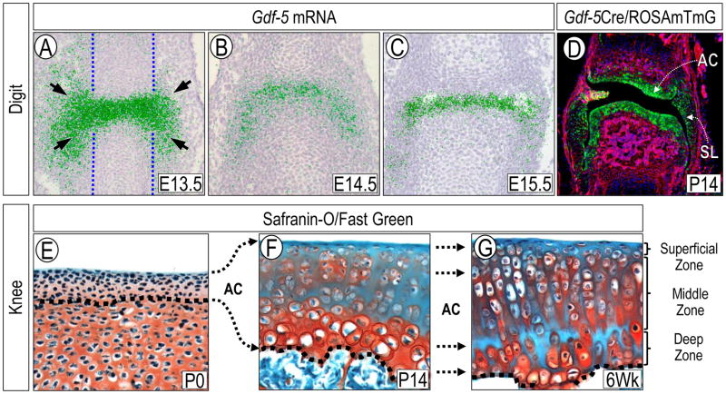
Articular cartilage has obvious and fundamental roles in joint function and body movement. Much is known about its organization, extracellular matrix, and phenotypic properties of its cells, but less is known about its developmental biology. Incipient articular cartilage in late embryos and neonates is a thin tissue with scanty matrix and small cells, while adult tissue is thick and zonal and contains large cells and abundant matrix. What remains unclear is not only how incipient articular cartilage forms, but how it then grows and matures into a functional, complex, and multifaceted structure. This review focuses on recent and exciting discoveries on the developmental biology and growth of articular cartilage, frames them within the context of classic studies, and points to lingering questions and research goals. Advances in this research area will have significant relevance to basic science, and also considerable translational value to design superior cartilage repair and regeneration strategies.
Keywords: Articular cartilage; Extracellular matrix; Joint disease; Joint formation; Lineage tracing; Progenitor cells.
Conflict of interest statement
The authors of this paper declare they have no conflicts of interest
Figures
References
-
- Hunziker E, Quinn T, Häuselmann H-J. Quantitative structural organization of normal adult human articular cartilage. Osteoarthr Cartil. 2002;10(7):564–572. - PubMed
-
- Mankin HJ. The reaction of articular cartilage to injury and osteoarthritis (first of two parts) N Engl J Med. 1974 Dec 12;291(24):1285–1292. - PubMed
-
- Dowthwaite GP, Bishop JC, Redman SN, et al. The surface of articular cartilage contains a progenitor cell population. J Cell Sci. 2004;117(6):889–897. - PubMed
Publication types
MeSH terms
Grants and funding
LinkOut - more resources
Full Text Sources
Other Literature Sources
