Review
. 2015 Oct;147(2):302-16.
doi: 10.1093/toxsci/kfv130.

Adaptive Posttranslational Control in Cellular Stress Response Pathways and Its Relationship to Toxicity Testing and Safety Assessment

Affiliations
Review

Adaptive Posttranslational Control in Cellular Stress Response Pathways and Its Relationship to Toxicity Testing and Safety Assessment

Qiang Zhang et al. Toxicol Sci. 2015 Oct.

Abstract

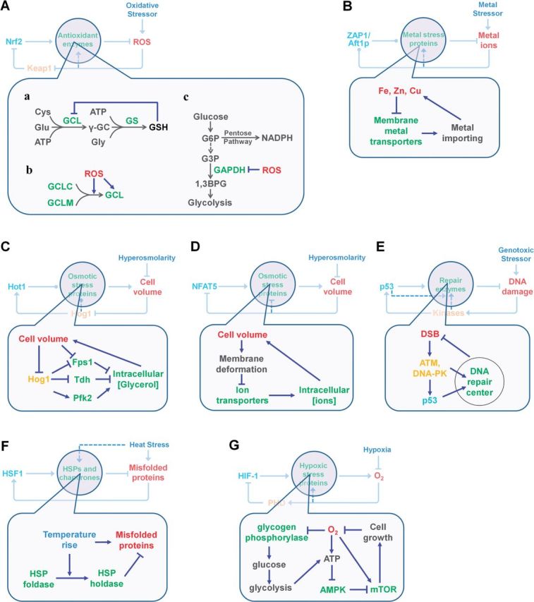
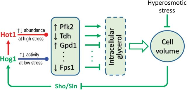
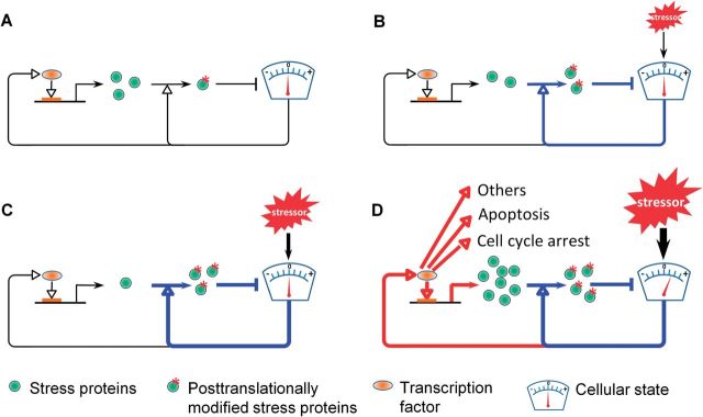
Although transcriptional induction of stress genes constitutes a major cellular defense program against a variety of stressors, posttranslational control directly regulating the activities of preexisting stress proteins provides a faster-acting alternative response. We propose that posttranslational control is a general adaptive mechanism operating in many stress pathways. Here with the aid of computational models, we first show that posttranslational control fulfills two roles: (1) handling small, transient stresses quickly and (2) stabilizing the negative feedback transcriptional network. We then review the posttranslational control pathways for major stress responses-oxidative stress, metal stress, hyperosmotic stress, DNA damage, heat shock, and hypoxia. Posttranslational regulation of stress protein activities occurs by reversible covalent modifications, allosteric or non-allosteric enzymatic regulations, and physically induced protein structural changes. Acting in feedback or feedforward networks, posttranslational control may establish a threshold level of cellular stress. Sub-threshold stresses are handled adequately by posttranslational control without invoking gene transcription. With supra-threshold stress levels, cellular homeostasis cannot be maintained and transcriptional induction of stress genes and other gene programs, eg, those regulating cell metabolism, proliferation, and apoptosis, takes place. The loss of homeostasis with consequent changes in cellular function may lead to adverse cellular outcomes. Overall, posttranslational and transcriptional control pathways constitute a stratified cellular defense system, handling stresses coherently across time and intensity. As cell-based assays become a focus for chemical testing anchored on toxicity pathways, examination of proteomic and metabolomic changes as a result of posttranslational control occurring in the absence of transcriptomic alterations deserves more attention.

Keywords: feedback; pathway; posttranslational; stress; transcriptional.


Figures

FIG. 1.
FIG. 1.
BMD evaluation and DRC formation in DNA damage response. A, BMD estimates for various in vitro endpoints in HT-1080 cells after 24-h treatments with 3 compounds respectively, ETP (etoposide), QUE (quercetin), and MMS (methylmethane sulfonate) capable of causing DNA damage—measured as micronuclei formation. Although activation of gene transcription program was expected to moderate stress responses and prevent micronuclei formation, micronuclei were nevertheless the most sensitive endpoint measured in relation to their BMD. The data are adapted from a published table (Clewell et al., 2014). B–D, Dynamic behaviors of DRCs in response to various levels of genotoxic damage by radiomimic chemical neocarzinostatin (NCS) in HT-1080 cells. B, Images of DRC foci detected by TP53BP1 antibody at various times following treatment with 5 or 25 ng/ml NCS. C, Dynamics of quantified TP53BP1 foci/cell in response to various concentrations of NCS treatment. Foci form quickly but only resolve relatively quickly with low NCS concentrations whereas high NCS concentrations lead to more persistent unresolved foci beyond 24 h with potential adverse cellular outcomes. D, TP53BP1 foci/cell remaining at 24 h in (B) (solid line, NOEL = 5 ng/ml) and cumulative (total) TP53BP1 foci/cell produced during the first 24 h (dashed line, NOEL = 0.5 ng/ml). The data were originally reported in Sun et al. (2014).
FIG. 2.
FIG. 2.
Cellular stress response to external stressor (S) may involve both transcriptional (outer solid lines) and posttranslational control (inner dotted lines) of the cellular state (Y). The transcriptional induction of stress genes (g) is activated by transcription factor (T) either through feedback (bottom arm) or feedforward (top arm). Posttranslational control bypasses the slow-acting transcriptional loops by regulating the activities of preexisting molecules of stress proteins (G) through covalent modifications or other fast mechanisms (dotted lines). Pointed arrows denote activation and blunted arrows denote inhibition.
FIG. 3.
FIG. 3.
Simulations comparing the different dynamic behaviors in the absence (A) versus presence (B) of posttranslational control. Posttranslational control allows effective handling of transient stresses. C, Transcriptionally mediated induction of the stress protein G cannot keep up with the rapidly changing stressor S, resulting in nearly unmitigated consequences on the cellular state variable, Y. D, Posttranslational activation of stress protein G results in more rapid upregulation of anti-stress activity, occurring almost in sync with the changing stressor S. The cellular state Y shows very brief increases at the initiation of the square wave for stressor S and then brief decreases with the cessation of the square wave input. Posttranslational control stabilizes the transcriptionally mediated negative feedback circuit, reducing pathway oscillations. E, A transcriptionally mediated negative feedback circuit with high amplification is prone to oscillation. F, Adding a fast-acting posttranslational pathway eliminates the time delay and damps out or eliminates oscillation. See Supplementary Material and the accompanying SBML model files for model details.
FIG. 4.
FIG. 4.
Posttranslational control processes in stress response pathways. A, Oxidative stress response, B, Yeast metal stress response, C, Yeast hyperosmotic stress response, D, Mammalian hyperosmotic stress response, E, DNA damage response, F, Heat shock response, and G, hypoxic response. Denotations of colored arrow heads: pointed, activation; blunted, inhibition; dotted, activation or inhibition. Refer to the text for details of these processes.
FIG. 5.
FIG. 5.
Schematic illustration of Hog1-mediated posttranslational and transcriptional hyperosmotic stress response pathway. At low stress levels, activated Hog1 posttranslationally alters the activities of a suite of enzymes involved in glycerol metabolism (Pfk2, Tdh, Gpd1, Fps1, etc.), resulting in increased production and decreased exportation of intracellular glycerol. At high stress levels, Hog1 activates transcription factor Hot1, which transcriptionally regulates the expression of the enzymes above, resulting in alterations in their abundance, which helps increase intracellular glycerol concentration on a longer time scale. Denotations of line colors: blue, posttranslational control; red, transcriptional control; green, common pathway shared by both posttranslational and transcriptional control. Denotations of colored arrow heads: pointed, activation; blunted, inhibition; dotted, activation or inhibition depending on target.
FIG. 6.
FIG. 6.
A proposed model for coherent transition from posttranslational control to transcriptional control as stressor level increases. A, At basal condition in the absence of exogenous stressor, a small fraction of preexisting stress proteins are posttranslationally modified to cope with background/endogenous stress. B, At very low stressor levels, more preexisting stress proteins are posttranslationally modified and thus activated to maintain homeostasis. C, At slightly higher stressor levels, even more preexisting stress proteins are posttranslationally modified and activated to maintain homeostasis. D, At considerably higher stressor levels, preexisting stress proteins are exhausted in terms of posttranslational modification; the cellular state cannot be maintained at the baseline level, and transcriptional induction of stress genes and genes responsible for cell cycle arrest, apoptosis, and other functional changes starts to occur. At these stressor levels, loss of cellular homeostasis and altered cellular function/fate may lead to adverse outcomes. The dial denotes the cellular state that is perturbed and needs to be maintained.

References

    1. Al Rashid S. T., Dellaire G., Cuddihy A., Jalali F., Vaid M., Coackley C., Folkard M., Xu Y., Chen B. P., Chen D. J., et al. (2005). Evidence for the direct binding of phosphorylated p53 to sites of DNA breaks in vivo. Cancer Res. 65, 10810–10821. - PubMed
    1. Anderson C. P., Shen M., Eisenstein R. S., Leibold E. A. (2012). Mammalian iron metabolism and its control by iron regulatory proteins. Biochim. Biophys. Acta 1823, 1468–1483. - PMC - PubMed
    1. Arsham A. M., Howell J. J., Simon M. C. (2003). A novel hypoxia-inducible factor-independent hypoxic response regulating mammalian target of rapamycin and its targets. J. Biol. Chem. 278, 29655–29660. - PubMed
    1. Backos D. S., Fritz K. S., Roede J. R., Petersen D. R., Franklin C. C. (2011). Posttranslational modification and regulation of glutamate-cysteine ligase by the alpha,beta-unsaturated aldehyde 4-hydroxy-2-nonenal. Free Radic. Biol. Med. 50, 14–26. - PMC - PubMed
    1. Beese S. E., Negishi T., Levin D. E. (2009). Identification of positive regulators of the yeast fps1 glycerol channel. PLoS Genet. 5, e1000738. - PMC - PubMed

Publication types

MeSH terms